. 2016 Dec 15;143(24):4665-4675.
doi: 10.1242/dev.144733. Epub 2016 Nov 8.

Nodal and FGF coordinate ascidian neural tube morphogenesis

Affiliations

Nodal and FGF coordinate ascidian neural tube morphogenesis

Ignacio A Navarrete et al. Development. .

Abstract

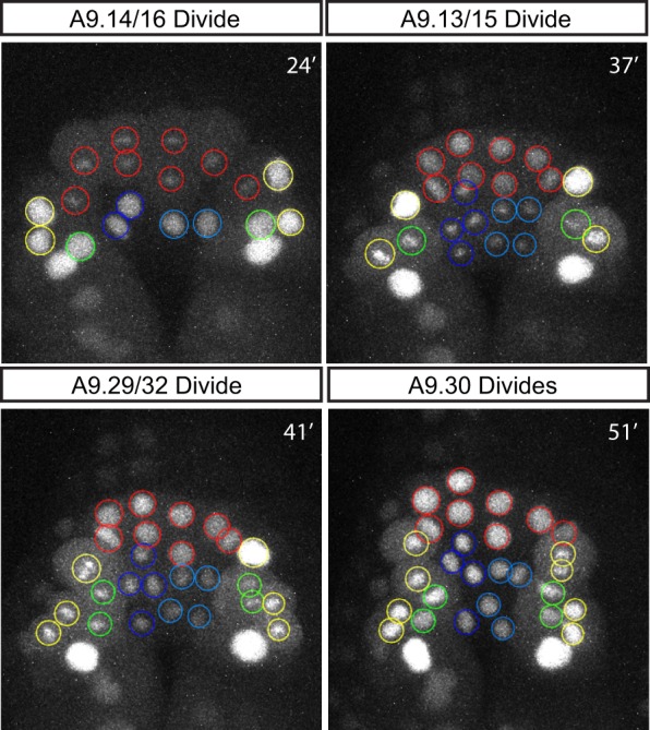
Formation of the vertebrate neural tube represents one of the premier examples of morphogenesis in animal development. Here, we investigate this process in the simple chordate Ciona intestinalis Previous studies have implicated Nodal and FGF signals in the specification of lateral and ventral neural progenitors. We show that these signals also control the detailed cellular behaviors underlying morphogenesis of the neural tube. Live-imaging experiments show that FGF controls the intercalary movements of ventral neural progenitors, whereas Nodal is essential for the characteristic stacking behavior of lateral cells. Ectopic activation of FGF signaling is sufficient to induce intercalary behaviors in cells that have not received Nodal. In the absence of FGF and Nodal, neural progenitors exhibit a default behavior of sequential cell divisions, and fail to undergo the intercalary and stacking behaviors essential for normal morphogenesis. Thus, cell specification events occurring prior to completion of gastrulation coordinate the morphogenetic movements underlying the organization of the neural tube.

Keywords: Ascidian; FGF; Intercalation; Morphogenesis; Neurulation; Nodal.


Conflict of interest statement

The authors declare no competing or financial interests.

Figures

Fig. 1.
Fig. 1.
Ciona A-line neural development. (A) Tail nerve cord lineages at mid-gastrula and mid-tailbud stages. Dark blue cells represent the A-lineage, which contributes to the ventral and lateral nerve cord; light blue represents b-line cells contributing to the dorsal nerve cord. Gray represents a-line neural cells at the mid-gastrula stage and the a-line-derived anterior sensory vesicle in tailbud embryo. Other tissues in the tailbud diagram are notochord (red), muscle (orange), endoderm (yellow) and epidermis (white). Lateral view of tailbud is a mid-sagittal section. Black bar shows location of tail cross-section. (B) Specification of A-line neural cells by Nodal and FGF signals. On the left side, blastomeres are labeled according to ascidian nomenclature. Colors represent A-line neural cell lineages (red, medial row II; yellow, lateral row II; blue, medial row I; green, lateral row I) and symbols represent signaling as shown in the key. A9.31 contributes to the tail muscles and is therefore uncolored. At the 44-cell stage, Nodal originating from the b6.5 blastomere signals to A7.8 but not A7.4. At the 110-cell stage an FGF signal of unknown origin is transduced, ultimately leading to MAPK activation in row I but not row II at the mid-gastrula stage.
Fig. 2.
Fig. 2.
Revised A-line neural lineage. (A-D,I) Time-lapse images of an embryo electroporated with FoxB>H2B:YFP and FoxB>lacZ from mid-gastrula stage to mid-tailbud stage. Circled cells belong to the A-line neural lineage. Cells were manually traced and labeled with Fiji trackmate plugin. Where cells from the left and right sides of the embryo mix, right-side cells are indicated by a dot within the nucleus. (E-H) False-colored images of phalloidin-stained embryos labeled with cell identities corresponding to the cells tracked in A-D. Embryos were electroporated with FoxB>H2B:Cherry to aid in cell identification and lacZ as a control for later experiments. (E) Three-dimensional projection of a mid-gastrula-stage embryo. Labeled cells are A-line of the ninth generation. (F-H) Single confocal slices from embryos at the indicated stages. (F) Labeled cells are of the tenth generation. (G) Labeled cells are of the tenth or 11th generation, as indicated. (H) Labeled cells are of the 11th generation. (J) Identities of A-line neural cells tracked to the mid-tailbud stage. Dashed circle represents the presumed location of A11.52, which moves beyond stack depth at 182 min. The following deviations from the published fate map were observed in this and other time-lapse experiments: A9.14 contributes to lateral walls (7/7), sixteen A9.29 derivatives at late neurula stage (6/6), anterior migration of A10.64 (3/3, unable to continuously trace in other experiments). Underlined labels indicate cells derived from right-side blastomeres. Colors reflect the lineage of each cell as in Fig. 1B. PSV, posterior sensory vesicle. Scale bars: 25 µm.
Fig. 3.
Fig. 3.
Timing of entry into the tenth generation. Stills from time-lapse imaging of a FoxB>H2B:YFP- and FoxB>lacZ-electroporated embryo (Movie 1) showing A-line cells entering the tenth generation at the indicated time points. In control embryos, medial row II cells always divide first (n=7/7) and A9.30 is always last (n=7/7). Colors reflect the lineage of each cell as in Fig. 1B.
Fig. 4.
Fig. 4.
FGF/MAPK is required for midline convergence of floor plate cells. (A-C) Time-lapse confocal microscopy of an embryo co-electroporated with FoxB>dnFGFR and FoxB>H2B:YFP. Cell lineages are color coded as in Fig. 1 and right-side nuclei at the midline are indicated with a spot. (B) Derivatives of A9.14, 16, 13 and 15 divide simultaneously (n=3/3). (C) At mid-neurula stage, most A9.13- and A9.15-derived cells have divided twice and failed to cross the midline (n=3/3). (D,E) False-colored phalloidin-stained mid-neurula embryos electroporated with FoxB>H2B:Cherry alone (D) or FoxB>dnFGFR together with FoxB>H2B:Cherry (E). In control embryos, A9.14 derivatives are confined to either side of the midline (arrow) whereas A9.13 and A9.15 derivatives converge, interrupting the membrane spanning the midline (arrowhead). In embryos electroporated with FoxB>dnFGFR, this midline span continues between left and right A9.13- and A9.15-derived cells (arrowhead). (F) Quantification of phenotypes for the indicated treatment. When convergence is normal, multiple floor plate cells are in contact with lateral cells from the left and right sides. Mild defects are defined by some rearrangement of floor plate cells with one or fewer cells spanning left and right lateral cells. Severe defects occur when floor plate cells are entirely confined to the side of the embryo where they originated; early divisions occur when one or more of these cells has entered the 11th generation by the mid-neurula stage. FoxB>H2B:Cherry total consists of 12 embryos co-electroporated with FoxB>lacZ and 13 embryos treated with DMSO at the 110-cell stage. Totals are from two independent experiments. Dashed line outlines A9.13 and A9.15 derivatives in the images in F. Scale bars: 25 µm.
Fig. 5.
Fig. 5.
FGF/MAPK acting through Ets/Elk family transcription factors delays cell division and promotes midline convergence of medial cells. (A-C) Time-lapse confocal microscopy of an embryo co-electroporated with FoxB>caMEK and FoxB>H2B:YFP. (B) Derivatives of A9.14, 16, 13 and 15 divide simultaneously (n=6/7). (C) At mid-neurula stage, A9.14 and A9.16 derivatives (red) have failed to enter the 11th generation and have converged towards the midline (n=5/7). (D) False-colored phalloidin-stained mid-neurula embryo electroporated with FoxB>caMEK and FoxB>H2B:Cherry. Arrowhead indicates where midline membrane is interrupted by A9.14 derivative. (E-G) Mid-neurula-stage embryos electroporated with the indicated transgenes and stained with phalloidin. Dashed line outlines A9.14/A9.16 derivatives. (E) Mnx reporter is limited to A9.13- and A9.15-derived floorplate cells. (F,G) Ectopic Mnx expression is seen in A9.14 and A9.16 derivatives. In both cases, these cells also interrupt the midline membrane (arrowheads). Floorplate cells are deeper within embryo. Number of embryos that exhibit ectopic Mnx reporter expression in combination with delayed division and midline convergence of A9.14/A9.16 derivatives is shown. Totals are from two independent experiments. (H) Quantification of A9.14 and A9.13 derivatives that have entered the 11th generation by the mid-neurula stage. Embryos were electroporated with FoxB driving the indicated gene and FoxB>H2B:Cherry to aid quantification. Totals are from two independent experiments. Scale bars: 25 µm.
Fig. 6.
Fig. 6.
Cell cycle delay is not sufficient for full midline convergence. (A) Quantification of A9.14 derivatives that have entered the 11th generation by the mid-neurula stage. Embryos were electroporated with FoxB driving the indicated gene and FoxB>H2B:Cherry to aid quantification. Aphidicolin treatment was carried out at the late gastrula stage. Total number of embryos counted from four independent experiments: lacZ: 16, lacZ+aphidicolin: 30, caMEK: 16, caMEK+aphidicolin: 28. (B) Box plot summary of midline membrane span length between A9.14 derivatives at mid-neurula stage. Measurements were performed manually with ImageJ software. Statistical significance calculated with Wilcoxon two-sample test. (C,D) Examples of membrane span traces (yellow lines) used to create the data shown in B. Embryos were electroporated with the indicated transgenes and treated with aphidicolin.
Fig. 7.
Fig. 7.
Junction rearrangement during midline convergence. Stills from time-lapse live imaging of embryos electroporated with the indicated transgenes. Numbers mark the same cell from one frame to the next with cell 3 inserting between cells 1 and 2 in both cases. Arrow indicates rearrangement of junction configuration between cells 1 and 2. Pseudo-color look-up table was chosen to increase membrane visibility. (A) Intercalation of medial row I cells. 1: A10.25 (left), 2: A10.26 (left), 3: A10.25 (right). (B) Intercalation of medial row II cells. 1: A10.27 (left), 2: A10.28 (left), 3: A10.27 (right). Scale bars: 10 µm.
Fig. 8.
Fig. 8.
Nodal influences timing of division and is required for proper lateral stacking during neurulation. Stills from time-lapse imaging visualizing H2B:YFP. Cell lineages are color coded as in Fig. 1 and right-side nuclei at the midline are indicated with a spot. (A-D) Embryo electroporated with FoxD>Lefty and Etr> H2B:YFP. (B,C) All cells belonging to row II divide simultaneously followed by all cells in row I (n=5/5). (D) Lateral stacking of A9.32, 30, 29 and 16 is disrupted and lateral row II cells have prematurely reached the 11th generation (n=6/6). (E,F) Embryo electroporated with FoxD>Nodal and Snail>H2B:YFP. (F) A9.14/16 cells (red) have failed to enter 11th generation by mid-neurula stage and lateral stacking of A9.32, 30, 29 and 16 is normal (n=3/3). Scale bars: 25 µm.
Fig. 9.
Fig. 9.
Stacking defects caused by Nodal inhibition. (A-D) Embryos electroporated with the indicated transgenes and fixed at the mid-neurula stage. (A,C) False-colored, phalloidin-stained embryos with n value indicating the number of embryos exhibiting four (A) or six (C) A-line cell columns. (B,D) Counting of floor plate cells. Dashed box indicates the same region of embryo as shown in A,C. Number of embryos with eight (B) or 12 (C) floor plate cells is indicated. Totals are from two independent experiments. Scale bars: 25 µm. (E) False-colored stills from time-lapse live imaging of embryos electroporated with FoxB>YFP-CAAX alone (left) or in combination with FoxD>Lefty (right). Arrows indicate junction between A9.30 and A9.32. Scale bars: 10 µm.
Fig. 10.
Fig. 10.
Oriented divisions are a morphogenetic ground state for the A-line neural lineage, which is modified by FGF/MAPK and Nodal signaling. (A-F) Stills from time-lapse live imaging of embryos electroporated with the indicated transgenes and FoxB>H2B:YFP reporter. Cell lineages are color coded as in Fig. 1B. Where cells from the left and right sides of the embryo mix, right side cells are indicated by a dot within the nucleus. (B,E) Late gastrula stage embryos exhibit similar phenotypes in the presence and absence of FGF/MAPK signaling. (C) Combined Nodal and FGF inhibition causes all A-line neural cells to enter the tenth generation within the same 15 min window (n=5/6) and enter 11th generation by mid-neurula stage without midline convergence or lateral stacking (n=6/6). (F) When MAPK signaling is activated in the absence of Nodal signaling all cells enter the tenth generation within the same 15 min window (n=3/6) and no cells enter the 11th generation by the mid-gastrula stage (n=4/7). Ectopic intercalations are present among both medial and lateral cells (n=4/7). (G) Visual summary of findings. Top row: At the late gastrula stage, cells have entered the tenth generation via an anterior-posterior oriented division and are positioned next to their sister cells. Midline is indicated by a dashed line. Bottom row: In the absence of earlier Nodal and FGF signals, cells divide again in an anterior-posterior orientation maintaining their position relative to the midline. When MAPK is activated in the absence of Nodal, cells delay their division and converge at the midline becoming separate from their sister cells. If cells have received a Nodal signal, entry into the 11th generation is delayed and cells form lateral stacks without losing contact with their tenth generation sisters. Opposing arrowheads between cells indicate the most recent cell division.

References

    1. Bertrand V., Hudson C., Caillol D., Popovici C. and Lemaire P. (2003). Neural tissue in ascidian embryos is induced by FGF9/16/20, acting via a combination of maternal GATA and Ets transcription factors. Cell 115, 615-627. 10.1016/S0092-8674(03)00928-0 - DOI - PubMed
    1. Böttcher R. T. and Niehrs C. (2005). Fibroblast growth factor signaling during early vertebrate development. Endocr. Rev. 26, 63-77. 10.1210/er.2003-0040 - DOI - PubMed
    1. Christiaen L., Wagner E., Shi W. and Levine M. (2009a). Isolation of sea squirt (Ciona) gametes, fertilization, dechorionation, and development. Cold Spring Harb. Protoc. 2009, pdb.prot5344. - PubMed
    1. Christiaen L., Wagner E., Shi W. and Levine M. (2009b). Electroporation of transgenic DNAs in the Sea Squirt Ciona. Cold Spring Harb. Protoc. 2009, pdb.prot5345. - PubMed
    1. Cole A. G. and Meinertzhagen I. A. (2004). The central nervous system of the ascidian larva: mitotic history of cells forming the neural tube in late embryonic Ciona intestinalis. Dev. Biol. 271, 239-262. 10.1016/j.ydbio.2004.04.001 - DOI - PubMed

Publication types

MeSH terms

Substances

LinkOut - more resources