

Dermatoscopic findings of pigmented purpuric dermatosis
- PMID: 27828629
- PMCID: PMC5087214
- DOI: 10.1590/abd1806-4841.20165124
Dermatoscopic findings of pigmented purpuric dermatosis
Abstract
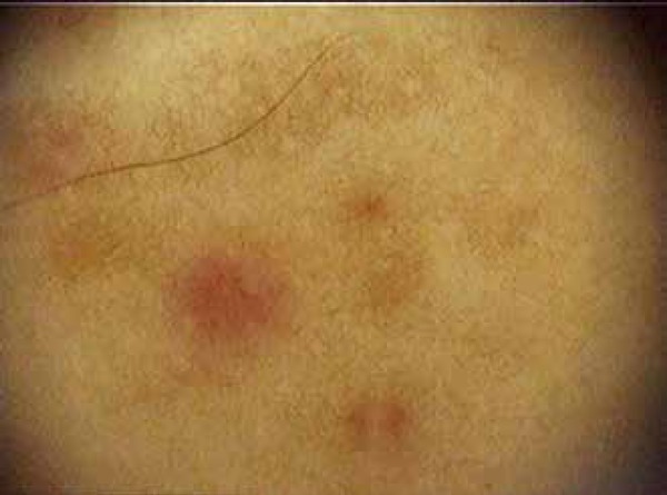
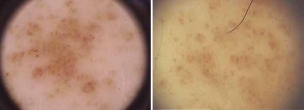
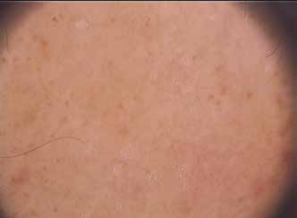
Background:: Pigmented purpuric dermatosis is a chronic skin disorder of unknown aetiology characterised by symmetrical petechial and pigmented macules, often confined to the lower limbs. The aetiology of pigmented purpuric dermatosis is unknown. Dermatoscopy is a non-invasive diagnostic technique that allows the visualisation of morphological features invisible to the naked eye; it combines a method that renders the corneal layer of the skin translucent with an optical system that magnifies the image projected onto the retina.
Objectives:: The aim of this study is to investigate the dermatoscopic findings of pigmented purpuric dermatosis.
Methods:: This study enrolled patients diagnosed histopathologically with pigmented purpuric dermatosis who had dermatoscopic records. We reviewed the dermatoscopic images of PPD patients who attended the outpatient clinic in the Istanbul Dermatovenereology Department at the Bezmialem Vakıf University Medical Faculty.
Results:: Dermatoscopy showed: coppery-red pigmentation (97%, n = 31) in the background, a brown network (34%, n = 11), linear vessels (22%, n = 7), round to oval red dots, globules, and patches (69%, n = 22; 75%, n = 24; 34%, n = 11; respectively), brown globules (26%, n = 8) and dots (53%, n = 17), linear brown lines (22%, n = 7), and follicular openings (13%, n = 4).
Conclusion:: To our knowledge, this is the first study to report the dermatoscopy of pigmented purpuric dermatosis. In our opinion, dermatoscopy can be useful in the diagnosis of pigmented purpuric dermatosis.
Conflict of interest statement
None
Figures
References
-
- Ehsani AH, Ghodsi SZ, Nourmohammad-Pour P, Aghazadeh N, Damavandi MR. Pigmented purpura dermatosis and viral hepatitis: A case control study. Australas J Dermatol. 2013;54:225–227. - PubMed
-
- Sardana K, Sarkar R, Sehgal VN. Pigmented purpuric dermatoses: An overview. Int J Dermatol. 2004;43:482–488. - PubMed
-
- Newton RC, Raimer SS. Pigmented purpuric eruptions. Dermatol Clin. 1985;3:165–169. - PubMed
-
- Taketuchi Y, Chinen T, Ichikawa Y, Ito M. Two cases of unilateral pigmented purpuric dermatosis. J Dermatol. 2001;28:493–498. - PubMed
-
- Martín JM, Bella-Navarro R, Jordá E. Vascular patterns in dermatoscopy. Actas Dermosifiliogr. 2012;103:357–375. - PubMed
MeSH terms
Substances
LinkOut - more resources
Full Text Sources
Other Literature Sources
Medical