. 2016 Nov 9;12(11):e1006429.
doi: 10.1371/journal.pgen.1006429. eCollection 2016 Nov.

Proteomic Landscape of Tissue-Specific Cyclin E Functions in Vivo

Affiliations

Proteomic Landscape of Tissue-Specific Cyclin E Functions in Vivo

Junko Odajima et al. PLoS Genet. .

Abstract

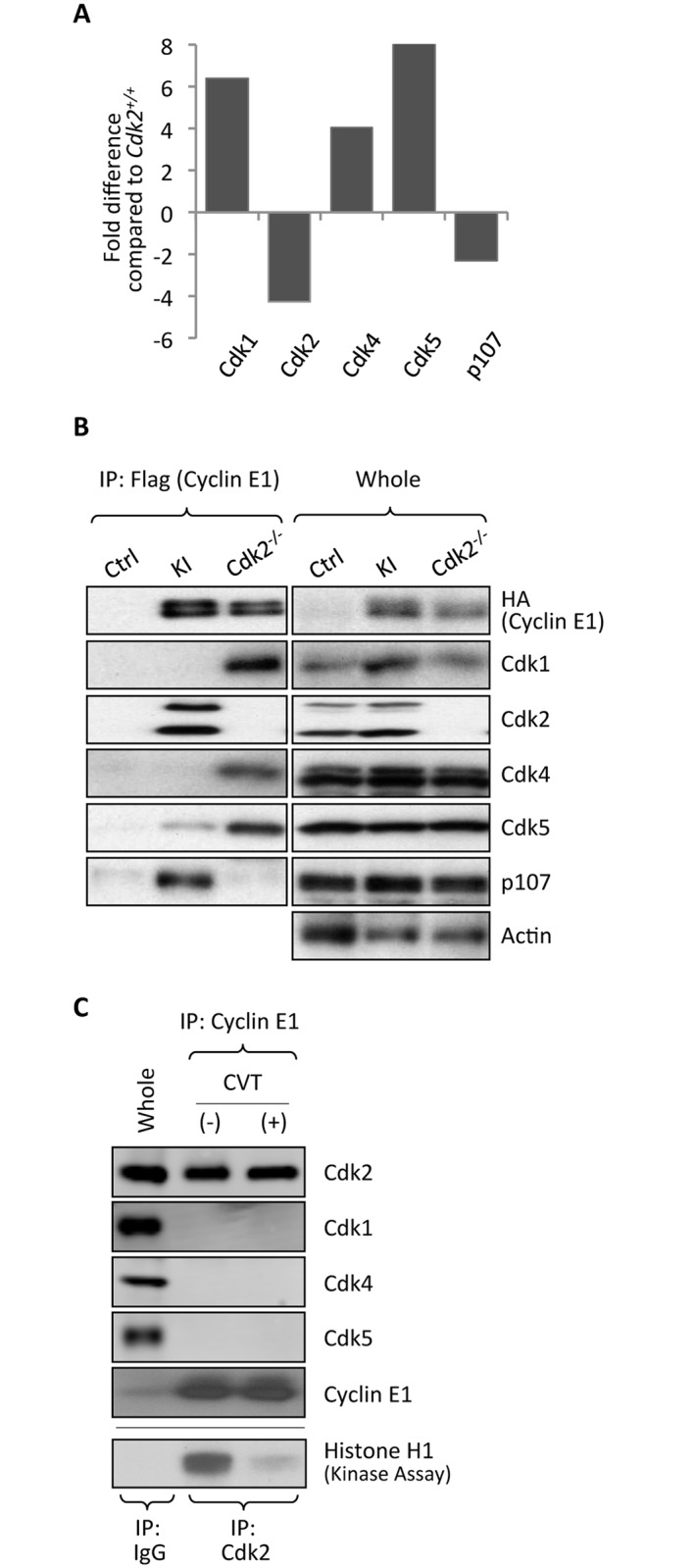
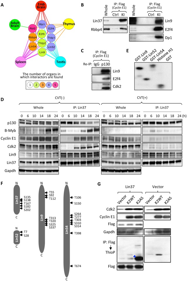
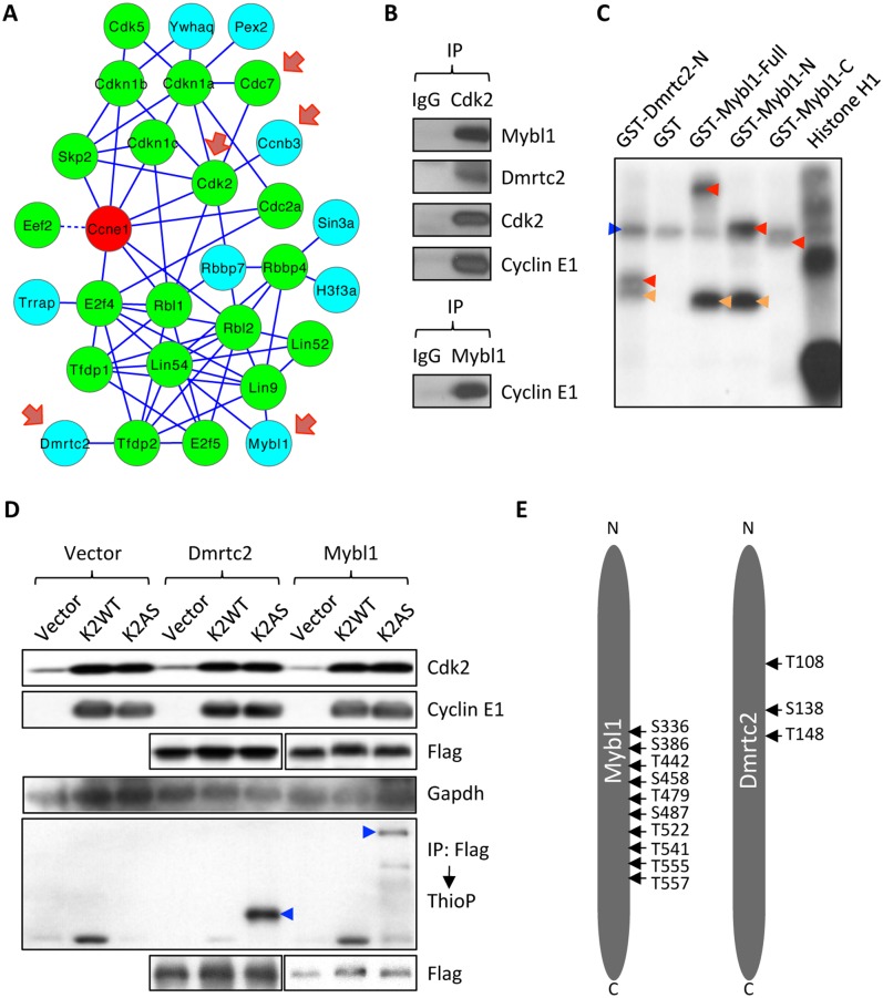
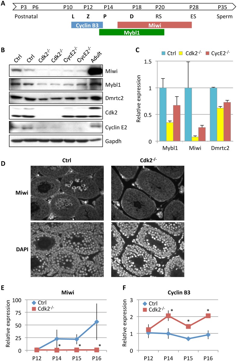
E-type cyclins (cyclins E1 and E2) are components of the cell cycle machinery that has been conserved from yeast to humans. The major function of E-type cyclins is to drive cell division. It is unknown whether in addition to their 'core' cell cycle functions, E-type cyclins also perform unique tissue-specific roles. Here, we applied high-throughput mass spectrometric analyses of mouse organs to define the repertoire of cyclin E protein partners in vivo. We found that cyclin E interacts with distinct sets of proteins in different compartments. These cyclin E interactors are highly enriched for phosphorylation targets of cyclin E and its catalytic partner, the cyclin-dependent kinase 2 (Cdk2). Among cyclin E interactors we identified several novel tissue-specific substrates of cyclin E-Cdk2 kinase. In proliferating compartments, cyclin E-Cdk2 phosphorylates Lin proteins within the DREAM complex. In the testes, cyclin E-Cdk2 phosphorylates Mybl1 and Dmrtc2, two meiotic transcription factors that represent key regulators of spermatogenesis. In embryonic and adult brains cyclin E interacts with proteins involved in neurogenesis, while in adult brains also with proteins regulating microtubule-based processes and microtubule cytoskeleton. We also used quantitative proteomics to demonstrate re-wiring of the cyclin E interactome upon ablation of Cdk2. This approach can be used to study how protein interactome changes during development or in any pathological state such as aging or cancer.


Conflict of interest statement

The authors have declared that no competing interests exist.

Figures

Fig 1
Fig 1. Generation of tagged cyclin E1 knock-In mice and analyses of cyclin E1-containing protein complexes.
(A and B) Targeting strategy to knock-in Flag and HA tags into the cyclin E1 locus to generate N-terminally tagged cyclin E1Ntag (A) and C-terminally tagged cyclin E1Ctag alleles (B). The exons are shown as green boxes, Flag tag as a blue box, and HA tag as a red box. Start and stop codons are marked with orange and yellow arrowheads, respectively. The hygromycin resistance cassette (Hyg) with flanking loxP sequences (filled arrows) is also indicated. Restriction enzyme recognition sites: E, EcoRI; A, AflII; Sc, ScaI; N, NotI; X, XhoI; K, KpnI; S, SpeI; P, PmeI; Hp, HpaI. Note that panel (A) was shown in ref [8]. (C) Western blot analysis of wild-type control (Ctrl), heterozygous cyclin E1+/Ntag, cyclin E1+/Ctag, and cyclin E1Ntag/Ctag embryonic stem cells probed with anti-cyclin E1 and -HA antibodies. Actin served as a loading control. Forth panel: cyclin E1 was immunoprecipitated with anti-Flag antibody and the immunoblots were probed with anti-Cdk2 antibody. Fifth panel: anti-Flag immunoprecipitates were used for in vitro kinase reactions using histone H1 as a substrate. (D) Same analyses as in (C) using spleens of homozygous knock-in mice. Lanes 1–2 in panels (C and D) were previously shown in [8]. (E) Cyclin E levels detected by western blotting in the indicated organs of 1-month-old mice and in embryonic brain (day E14.5). Actin served as a loading control. The last two lanes (Brain) were previously shown in [8]. (F) Quantification of cyclin E levels in different organs, normalized against actin (from E). (G) Protein lysates from brains and testes of adult tagged cyclin E1 knock-in mice were separated by gel-filtration chromatography. Fractions containing protein complexes of the indicated molecular weights were analyzed by western blotting for cyclin E using an anti-HA antibody. (H) Cyclin E1-associated proteins were purified from the indicated organs of tagged cyclin E1 knock-in (KI) mice, or from control mice (Ctrl, ‘mock’ purifications) by sequential immunoaffinity purifications with anti-Flag and -HA antibodies, and 10% of the final eluate was resolved on PAGE gels and silver-stained. Arrows indicate bands corresponding to cyclin E1. Panels representing embryonic and adult brains were previously shown in [8].
Fig 2
Fig 2. Cyclin E1-interactomes.
(A) Diagrams depicting cyclin E1-interacting proteins in the indicated mouse organs. Cyclin E1 is shown as a red node. Green nodes denote highest-confidence ‘core’ interactors (Category 1, see S1 Appendix). Yellow and blue nodes represent, respectively, lower confidence Categories 2 and 3 interactors that were included to the interactome based on their reported interaction with core interactors in the STRING database (see S1 Appendix). Solid lines depict STRING-verified interactions. Dashed lines depict an interaction derived from our mass spectrometry analyses between cyclin E1 and a protein that has no known interactions with other core interactors. (B) A combined diagram depicting cyclin E1-interacting proteins from all five organs analyzed. Cyclin E1 is shown as a red node. Green nodes denote highest-confidence core (Category 1) interactors. Yellow and blue nodes denote, respectively, Categories 2 and 3 interactors, which were included into the interactome based on their ability to interact with core interactors as revealed by STRING (see S1 Appendix). Solid blue lines depict STRING-verified interactions between pairs of proteins that were identified by us as cyclin E1-interacting proteins within the same organ. Gray dotted lines depict STRING-verified interactions between pairs of proteins identified as cyclin E1-interators in different organs. Blue dashed lines depict interactions detected in our mass spectrometry analyses between cyclin E1 and a protein that has no known interactions with other core proteins within the same organ interactome.
Fig 3
Fig 3. Analyses of cyclin E1-interactomes.
(A) Venn diagram depicting the numbers of unique and shared cyclin E1 interactors in the indicated organs. (B) Fraction of unique interactors in the indicated organs. (C) Pairwise comparisons of the fraction of cyclin E1-interactors shared between the indicated organs. (D) The fraction of cyclin E1 interactors in the indicated organs that were assigned to a given Gene Ontology category (see S2 Table). Categories assigned at least 10% of the interactors in a given organ are marked in red. CC, cell cycle; TX, transcription; Neuro, neuronal function; Cyto, microtubules/cytoskeleton; Ubiq, ubiquitination; Metab, metabolism. (E) Heatmap displaying functional enrichment of cyclin E1 interactors in Gene Ontology classes of biological processes. The five columns correspond to the five organs analyzed, and each horizontal row denotes a distinct biological process. Colors depict fold-enrichment for the cyclin E1 interactors from the particular organ in a given biological process, between green (fold-enrichment one or lower) to red (fold-enrichment five or higher). Only categories in which at least one organ had an EASE score of 0.2 or lower are shown (see S1 Appendix). Left panel shows a complete heatmap, right panels show selected common and organ-specific biological processes: cell cycle (red box, enriched in all organs), neurogenesis and synaptic plasticity (blue box, shared between embryonic and adult brains) and regulation of microtubule-based processes and microtubule cytoskeleton (green box, specific to adult brain).
Fig 4
Fig 4. Quantitative proteomic (iTRAQ) analysis of cyclin E1-interacting proteins in mouse organs in the absence of Cdk2.
(A) Relative abundance of cyclin E1-associated Cdk1, Cdk2, Cdk4, Cdk5 and p107 in the spleens of Cdk2-/-/cyclin E1Ntag/Ntag mice, as compared to Cdk2+/+/cyclin E1Ntag/Ntag animals, was determined by iTRAQ labeling and LC-MS. (B) The amount of cyclin E1-associated Cdk1, Cdk2, Cdk4, Cdk5 and p107 in the spleens of wild-type (Ctrl), Cdk2+/+/cyclin E1Ntag/Ntag (KI), and Cdk2-/-/cyclin E1Ntag/Ntag (Cdk2-/-) mice was gauged by immunoprecipitation with an anti-Flag antibody and immunoblotting with the indicated antibodies. Abundance of each protein in total lysates (whole) is also shown. (C) Spleens from wild-type mice were incubated with 20 μM CVT-313 (+) or with vehicle only (-). Association of cyclin E1 with Cdk2, Cdk1, Cdk4 and Cdk5 was assessed by IP–western blotting. Whole, whole cell lysate from vehicle only-treated mice. Lower panel: To ensure that CVT-313 treatment inhibited Cdk2 kinase activity, Cdk2 was immunoprecipitated from lysates and used for in vitro kinase reactions with histone H1 as a substrate. Note that CVT-313 treatment strongly decreased Cdk2 kinase activity.
Fig 5
Fig 5. Interaction of cyclin E1 with the DREAM complex.
(A) A diagram showing in which organ a particular component of the DREAM complex was identified as a cyclin E1-interacting protein by our mass spectrometric analyses. The number of organs in which a given protein was found to associate with cyclin E1 is depicted by the color. (B) Association of cyclin E1 with components of the DREAM complex in the spleens of tagged knock-in mice (KI) was verified by immunoprecipitating (IP) cyclin E1 with anti-Flag antibody followed by immunoblotting with the indicated antibodies. (C) IP followed by re-IP-immunoblotting to demonstrate that cyclin E1, Cdk2 and the DREAM complex components are present within the same multi-protein complex. Cyclin E1 was immunoprecipitated from spleens of KI mice using anti-Flag antibody, protein complexes were eluted with Flag peptides, re-immunoprecipitated with IgG (control) or with anti-p130 antibody, and then immunoblotted with the indicated antibodies. (D) T98G cells were serum starved for 72 hrs (0% FBS). Subsequently, cells were stimulated to re-enter the cell cycle by addition of 20% FBS supplemented with either 0.2% DMSO (control, left two panels) or 20 μM CVT-313 (right two panels), and harvested at the indicated time-points. Cell extracts (whole) as well as anti-Lin37 immunoprecipitates were resolved on 4–15% gradient SDS-PAGE gels and probed with indicated antibodies. Gapdh serves as a loading control. (E) Cyclin E1-Cdk2 kinase can phosphorylate purified recombinant Lin proteins in vitro. Lin9, Lin52 and Lin54 were expressed as GST-fusion proteins in E. Coli, purified and subjected to in vitro kinase reactions with the recombinant cyclin E1-Cdk2 in the presence of [γ32P]ATP. Recombinant histone H1 was used as a positive control and GST as a negative control. (F) A diagram illustrating amino acid residues in human Lin proteins that were phosphorylated by cyclin E-Cdk2. (G) Wild-type Cdk2 (K2WT) or analog-sensitive Cdk2 (K2AS) were transfected into 293T cells together with cyclin E1 and Flag-tagged Lin37 or vector control. After supplementing cells with 6-Fu-ATPγS, labeling of Lin37 was evaluated by immunoprecipitating Lin37 with an anti-Flag antibody followed by immunoblotting with anti-thiophosphate ester antibody. A blue arrowhead indicates 6-Fu-ATPγS-labeled Lin37.
Fig 6
Fig 6. Identification of Mybl1 and Dmrtc2 as cyclin E-Cdk2 phosphorylation substrates in the testes.
(A) A diagram illustrating cyclin E1-interactome in testes, consisting of highest-confidence ‘core’ interactors (green nodes), and lower-confidence Category 3 interactors (blue nodes) that were included to the interactome based on their reported interaction with core interactors in the STRING database (see S1 Appendix). Solid lines depict STRING-verified interactions. Dashed lines depict an interaction derived from our mass spectrometry analyses between cyclin E1 and a protein that has no known interactions with other ‘core’ interactors. Red arrows indicate proteins that were previously implicated to play important roles in spermatogenesis. (B) Interaction between endogenous Cdk2/cyclin E and Mybl1 and Dmrtc2 in mouse testes, detected by IP–western blotting. Cdk2 or Mybl1 were immunoprecipitated from lysates of testes, and immunoblots were probed with the indicated antibodies. (C) N-terminal fragment (aa 1–201) of Dmrtc2, as well as full length, N-terminal (aa 1–376), and C-terminal (aa 376–752) fragments of Mybl1 were expressed as GST-fusion proteins in E. Coli, purified and subjected to in vitro kinase reactions with the recombinant cyclin E1-Cdk2 in the presence of [γ 32P]ATP. Recombinant histone H1 was used as a positive control and GST as a negative control. Red arrowheads point to phosphorylated GST-fusion proteins, orange arrowheads indicate phosphorylated truncated proteins, and blue arrow indicates auto-phosphorylated recombinant cyclin E1-Cdk2. (D) Wild-type Cdk2 (K2WT) or analog-sensitive Cdk2 (K2AS) were transfected into 293T cells together with cyclin E1 and Flag-tagged substrates (Mybl1 or Dmrtc2). After supplementing cells with 6-Fu-ATPγS, labeling of substrates was evaluated by immunoprecipitating Mybl1 or Dmrtc2 with anti-Flag antibody followed by immunoblotting with an anti-thiophosphate ester antibody. Blue arrowheads indicate ATPγS-labeled Mybl1 and Dmrtc2. The experiment was performed on the same gel as the one shown in Fig 5G, hence the vector control (Vector) is identical. (E) A diagram illustrating amino acid residues in Mybl1 and Dmrtc2, which were phosphorylated by cyclin E-Cdk2.
Fig 7
Fig 7. Reduced levels of Mybl1 and Dmrtc2 and altered levels of Mybl1 transcriptional targets in Cdk2- and cyclin E-deficient testes.
(A) Schematic representation of expression patterns for Mybl1, Miwi, and cyclin B3 during spermatogenesis. White arrow shows progression of mouse spermatogenesis after birth (P, postnatal days). Meiotic phases (L, leptotene; Z, zygotene; P, pachytene; D, diplotene) are indicated in bold. RS, round spermatids; ES, elongating spermatids. (B) Levels of Miwi, Mybl1 and Dmrtc2 in the testes of P15 wild-type (Ctrl), Cdk2-/- and cyclin E2-/- mice were detected by immunoblotting. Protein lysates from testes of 1-month-old wild-type mice were used as a positive control (Adult). (C) Quantification of Mybl1, Miwi and Dmrtc2 levels shown in (B). Data were normalized to Ctrl and represent mean ± SD. (D) Sections of seminiferous tubules obtained from testes of P15 wild-type (Ctrl) and Cdk2-/- mice were stained with anti-Miwi antibody followed by Alexa 568. Lower panels: DAPI staining to visualize cell nuclei. (E and F) RT-qPCR analyses to gauge levels of Miwi (E) and cyclin B3 (F) transcripts at the indicated postnatal days (P12–P16) in the testes of wild-type (Ctrl) or Cdk2-/- mice. Error bars represent SD (n = 4 for Ctrl; n = 5 for Cdk2-/- at each time-point). *p<0.05 using unpaired t test.

Similar articles

Cited by

References

    1. Hwang HC, Clurman BE (2005) Cyclin E in normal and neoplastic cell cycles. Oncogene 24: 2776–2786. 10.1038/sj.onc.1208613 - DOI - PubMed
    1. Satyanarayana A, Kaldis P (2009) Mammalian cell-cycle regulation: several Cdks, numerous cyclins and diverse compensatory mechanisms. Oncogene 28: 2925–2939. 10.1038/onc.2009.170 - DOI - PubMed
    1. Geng Y, Yu Q, Sicinska E, Das M, Schneider JE, et al. (2003) Cyclin E ablation in the mouse. Cell 114: 431–443. - PubMed
    1. Parisi T, Beck AR, Rougier N, McNeil T, Lucian L, et al. (2003) Cyclins E1 and E2 are required for endoreplication in placental trophoblast giant cells. Embo J 22: 4794–4803. 10.1093/emboj/cdg482 - DOI - PMC - PubMed
    1. Martinerie L, Manterola M, Chung SS, Panigrahi SK, Weisbach M, et al. (2014) Mammalian E-type cyclins control chromosome pairing, telomere stability and CDK2 localization in male meiosis. PLoS Genet 10: e1004165 10.1371/journal.pgen.1004165 - DOI - PMC - PubMed

MeSH terms