. 2016 Nov 9;11(11):e0165412.
doi: 10.1371/journal.pone.0165412. eCollection 2016.

Remarkable Diversity and Prevalence of Dagger Nematodes of the Genus Xiphinema Cobb, 1913 (Nematoda: Longidoridae) in Olives Revealed by Integrative Approaches

Affiliations

Remarkable Diversity and Prevalence of Dagger Nematodes of the Genus Xiphinema Cobb, 1913 (Nematoda: Longidoridae) in Olives Revealed by Integrative Approaches

Antonio Archidona-Yuste et al. PLoS One. .

Abstract

The genus Xiphinema includes a remarkable group of invertebrates of the phylum Nematoda comprising ectoparasitic animals of many wild and cultivated plants. Damage is caused by direct feeding on root cells and by vectoring nepoviruses that cause diseases on several crops. Precise identification of Xiphinema species is critical for launching appropriate control measures. We make available the first detailed information on the diversity and distribution of Xiphinema species infesting wild and cultivated olive in a wide-region in southern Spain that included 211 locations from which 453 sampling sites were analyzed. The present study identified thirty-two Xiphinema spp. in the rhizosphere of olive trees, ten species belonging to Xiphinema americanum-group, whereas twenty-two were attributed to Xiphinema non-americanum-group. These results increase our current knowledge on the biodiversity of Xiphinema species identified in olives and include the description of four new species (Xiphinema andalusiense sp. nov., Xiphinema celtiense sp. nov., Xiphinema iznajarense sp. nov., and Xiphinema mengibarense sp. nov.), and two new records for cultivate olives (X. cadavalense and X. conurum). We also found evidence of remarkable prevalence of Xiphinema spp. in olive trees, viz. 85.0% (385 out of 453 sampling sites), and they were widely distributed in both wild and cultivated olives, with 26 and 17 Xiphinema spp., respectively. Diversity indexes (Richness, Hill´s diversity, Hill´s reciprocal of D and Hill´s evenness) were significantly affected by olive type. We also developed a comparative morphological and morphometrical study together with molecular data from three nuclear ribosomal RNA genes (D2-D3 expansion segments of 28S, ITS1, and partial 18S). Molecular characterization and phylogenetic analyses allowed the delimitation and discrimination of four new species of the genus described herein and three known species. Phylogenetic analyses of Xiphinema spp. resulted in a general consensus of these species groups. This study is the most complete phylogenetic analysis for Xiphinema non-americanum-group species to date.


Conflict of interest statement

The authors have declared that no competing interests exist.

Figures
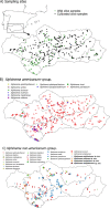
Fig 1
Fig 1. Geographic distribution of dagger nematodes of the genus Xiphinema in the present fieldworks on wild and cultivated olive in southern Spain.
This map may be similar but not identical to other published maps of Andalusia and is therefore for illustrative purposes only on the sampling sites.
Fig 2
Fig 2. Summary barplot of nematode abundance, Richness, Hill´s diversity (Hill´s 1), Hill´s reciprocal of D (Simpson´s dominance index) (Hill´s 2) and Hill´s evenness diversity indexes derived from results of Xiphinema spp. identification in 385 sampling sites of olives orchards (Fig 1) grouped by olive type (wild and cultivated olive) and X. americanum-group and X. non-americanum-group species.
Error bars indicate the standard error of the mean. Significance = F probability of main effects in ANOVA, according to Tukey´s test [49] for P < 0.05.
Fig 3
Fig 3
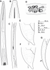
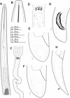
Line drawings of Xiphinema andalusiense sp. nov., female paratypes and first-stage juvenile A) Pharyngeal region. B) Detail of lip region. C) Posterior female genital branch showing Z-differentiation. D) Detail of Z-differentiation. E-F) Female tails. G) First-stage juvenile tail (J1).
Fig 4
Fig 4
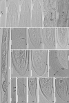
Light micrographs of Xiphinema andalusiense sp. nov., female paratypes and juvenile stages A) Pharyngeal region. B–E) Female anterior regions. F) Detail of anterior female gonad showing Z-differentiation. G) Vulval region. H) Detail of female genital track showing Z-differentiation. I-K) Z-differentiation. L-T) Female tails. U-X) First-, second-, third-, and fourth-stage juvenile (J1-J4) tails, respectively. Abbreviations: a = anus; cb = crystalloid bodies; gr = guiding-ring; odt = odontostyle; rodt = replacement odontostyle; spi = spiniform structures; spZ = Z-differentiation; v = vulva. Scale bars = 20 μm.
Fig 5
Fig 5
Relationship between body length and functional and replacement odontostyle (Ost and rOst, respectively) length in all developmental stages from first-stage juveniles (J1) to mature females of: A) Xiphinema andalusiense sp. nov. B) Xiphinema celtiense sp. nov. C) Xiphinema iznajarense sp. nov. D) Xiphinema mengibarense sp. nov.
Fig 6
Fig 6
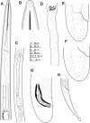
Line drawings of Xiphinema celtiense sp. nov., female paratypes, male and first-stage juvenile A) Pharyngeal region. B) Detail of lip region. C) Posterior female genital branch showing Z-differentiation. D) Detail of Z-differentiation. E-F) Female tails. G) Male tail. H) First-stage juvenile tail (J1).
Fig 7
Fig 7
Light micrographs of Xiphinema celtiense sp. nov., female paratypes, male and juvenile stages A) Pharyngeal region. B–D) Female anterior regions. E) Detail of female genital track showing Z-differentiation. F) Detail of anterior female gonad showing Z-differentiation. G) Z-differentiation. H-M) Female tails. N) Detail of first-stage anterior region. O-R) First-, second-, third-, and fourth-stage juvenile (J1-J4) tails, respectively. S) Male tail with detail of spicules. Abbreviations: a = anus; cb = crystalloid bodies; gr = guiding-ring; odt = odontostyle; rodt = replacement odontostyle; sp = spicules; spZ = Z-differentiation; v = vulva. Scale bars = 20 μm.
Fig 8
Fig 8
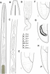
Line drawings of Xiphinema iznajarense sp. nov., female paratypes, male and first-stage juvenile A) Pharyngeal region. B) Detail of lip region. C) Anterior female genital branch showing Z-differentiation. D) Detail of Z-differentiation. E-F) Female tails. G) Male tail. H) First-stage juvenile tail (J1).
Fig 9
Fig 9
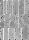
Light micrographs of Xiphinema iznajarense sp. nov., female paratypes, male and juvenile stages A-D) Female anterior regions. E) Detail of anterior female gonad. F) Vulval region. G-H) Detail of female genital track showing Z-differentiation. I-M) Female tails. N) Male tail with detail of spicules. P) Detail of first-stage anterior region. Q-T) First-, second-, third-, and fourth-stage juvenile (J1-J4) tails, respectively. Abbreviations: a = anus; cb = crystalloid bodies; gr = guiding-ring; odt = odontostyle; rodt = replacement odontostyle; sp = spicules; spi = spiniform structures; spl = ventromedian supplements; spZ = Z-differentiation; v = vulva. Scale bars = 20 μm.
Fig 10
Fig 10. Line drawings of Xiphinema mengibarense sp. nov., female paratypes, male and first-stage juvenile.
A) Pharyngeal region. B) Detail of lip region. C,D) Detail of Z-differentiation. E,F) Female tails. G) Male tail. H) First-stage juvenile tail (J1).
Fig 11
Fig 11
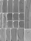
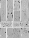
Light micrographs of Xiphinema mengibarense sp. nov., female paratypes, male and juvenile stages A-E) Female anterior regions. F-H) Detail of female genital track showing Z-differentiation. I-L) Female tails. M) Detail of male genital track showing sperm cells. N-O) Male tail with detail of spicules and ventromedian supplements. P) Detail of first-stage anterior region. Q-T) First-, second-, third-, and fourth-stage juvenile (J1-J4) tails, respectively. Abbreviations: a = anus; cb = crystalloid bodies; gr = guiding-ring; odt = odontostyle; rodt = replacement odontostyle; sp = spicules; spe = sperm cells; spi = spiniform structures; spl = ventromedian supplements; sss = spZ = Z-differentiation; v = vulva. Scale bars = 20 μm.
Fig 12
Fig 12
Light micrographs of Xiphinema cadavalense Bravo & Roca, 1995 females from Spain A) Neck region. B-D) Female lip regions. E) Vulval region. F-I) Details of pseudo-Z organ. J-M) Female tails. Abbreviations: a = anus; af = amphidial fovea; psZ = pseudo-Z organ. Scale bars = 20 μm.
Fig 13
Fig 13
Light micrographs of Xiphinema conurum Siddiqi, 1964 females from Spain A) Female lip region. B) Female anterior region showing detail of odontophore and flanges. C) Detail of female genital track showing Z-differentiation. D-E) Female tails. Abbreviations: a = anus; sk = flanges; sp = spiniform structures; spZ = Z-differentiation. Scale bars = 20 μm.
Fig 14
Fig 14
Light micrographs of Xiphinema pseudocoxi Sturhan, 1984, females from Spain A) Neck region. B-D) Details of lip region. E) Detail of pseudo-Z organ. F-K) Female tails showing morphological variability. Abbreviations: a = anus; gr = guiding ring; psZ = pseudo-Z organ. Scale bars A = 50 μm; B-K = 20 μm.
Fig 15
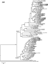
Fig 15. The 50% majority rule consensus tree from Bayesian inference analysis generated from the D2-D3 of 28S rRNA gene dataset of Xiphinema spp. with the GTR+I+G model.
Posterior probabilities more than 0.70 are given for appropriate clades. Newly obtained sequences are in bold letters. Scale bar = expected changes per site.
Fig 16
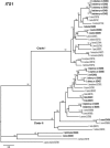
Fig 16. The 50% majority rule consensus trees from Bayesian inference analysis generated from the ITS rRNA gene dataset of Xiphinema spp. with the GTR+I+G model.
Posterior probabilities more than 0.70 are given for appropriate clades. Newly obtained sequences are in bold letters. Scale bar = expected changes per site.
Fig 17
Fig 17. The 50% majority rule consensus trees from Bayesian inference analysis generated from the partial 18S rRNA gene dataset of Xiphinema spp. with the TIM3+I+G model.
Posterior probabilities more than 0.70 are given for appropriate clades. Newly obtained sequences are in bold letters. Scale bar = expected changes per site.

References

    1. Wolters V. Biodiversity of soil animals and its function. European Journal of Soil Biology. 2001;37:221–7.
    1. Decaens T, Jiménez JJ, Gioia C, Measey GJ, Lavelle P. The values of soil animals for conservation biology. European Journal of Soil Biology. 2006;42:23–8.
    1. Bongers T, Ferris H. Nematode community structure as a bioindicator in environmental monitoring. Trends in Ecology and Evolution. 1999;14:224–8. - PubMed
    1. Wyss U. Root parasitic nematodes: an overview In: FMWG C. Fenoll, Ohl S.A., editor. Cellular and molecularaspects of plant-nematode interactions. 10. Dordrecht, The Netherlands: Kluwer Academic Publisher; 1997. p. 5–22.
    1. Decraemer W, Hunt D. Structure and clasiffication In: Perry RN, Moens M., editor. Plant Nematology. Wallingford, UK: CABI; 2006. p. 3–32.

LinkOut - more resources