. 2016 Nov 29;7(48):79869-79884.
doi: 10.18632/oncotarget.13084.

RSK2 activity mediates glioblastoma invasiveness and is a potential target for new therapeutics

Affiliations

RSK2 activity mediates glioblastoma invasiveness and is a potential target for new therapeutics

Florian J Sulzmaier et al. Oncotarget. .

Abstract

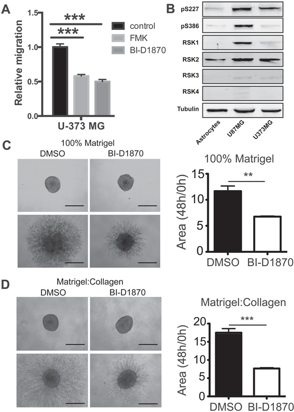
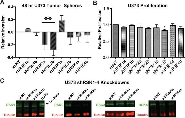
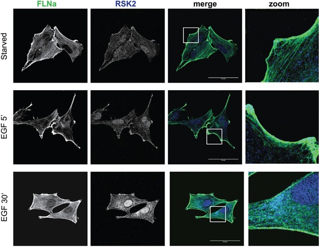
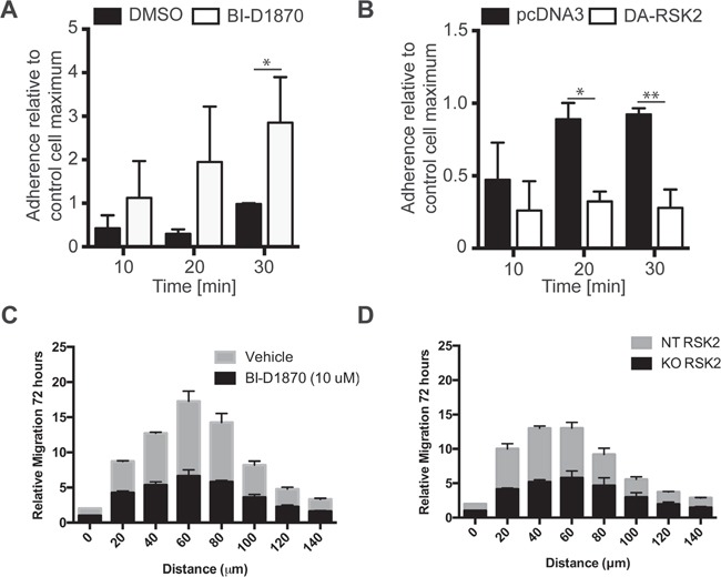
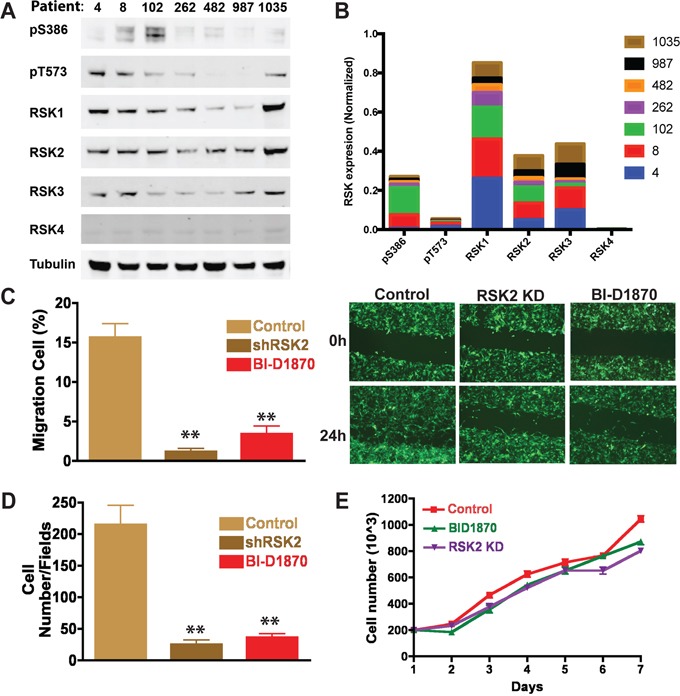
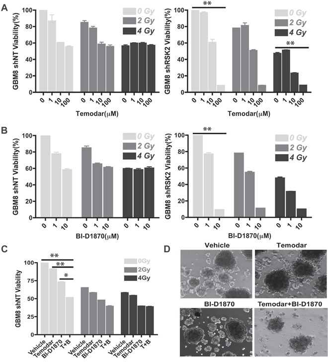
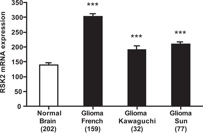
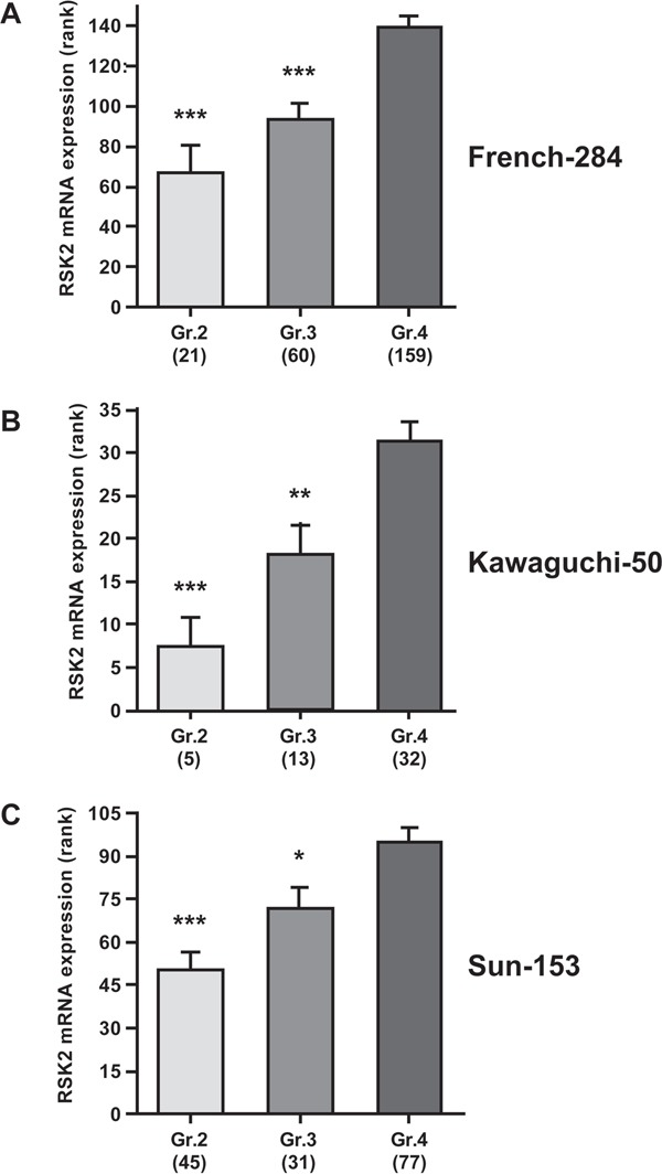
In glioblastoma (GBM), infiltration of primary tumor cells into the normal tissue and dispersal throughout the brain is a central challenge to successful treatment that remains unmet. Indeed, patients respond poorly to the current therapies of tumor resection followed by chemotherapy with radiotherapy and have only a 16-month median survival. It is therefore imperative to develop novel therapies. RSK2 is a kinase that regulates proliferation and adhesion and can promote metastasis. We demonstrate that active RSK2 regulates GBM cell adhesion and is essential for cell motility and invasion of patient-derived GBM neurospheres. RSK2 control of adhesion and migration is mediated in part by its effects on integrin-Filamin A complexes. Importantly, inhibition of RSK2 by either RSK inhibitors or shRNA silencing impairs invasion and combining RSK2 inhibitors with temozolomide improves efficacy in vitro. In agreement with the in vitro data, using public datasets, we find that RSK2 is significantly upregulated in vivo in human GBM patient tumors, and that high RSK2 expression significantly correlates with advanced tumor stage and poor patient survival. Together, our data provide strong evidence that RSK inhibitors could enhance the effectiveness of existing GBM treatment, and support RSK2 targeting as a promising approach for novel GBM therapy.

Keywords: ERK; GBM; MAPK; RSK; glioblastoma; invasion.


Conflict of interest statement

CONFLICTS OF INTEREST

The authors declare that they have no competing financial interests.

Figures

Figure 1
Figure 1. RSK isoforms are required for GBM migration and invasion
A. Migration of U-373 cells was determined in the presence of RSK inhibitors (FMK and BID1870) or control DMSO. Relative migration into the scratch was measured at 24 hours. B. Immunoblot showing expression of RSK1-4 isoforms in the indicated cells. C. Day 4 U-373MG tumor spheroids were embedded in either 100% matrigel or D. a 50% Matrigel/50% collagen mixture (right panel) and treated with DMSO or 10 μM BI-D1870. Images were acquired at 0, 24, and 48 hours after addition of drug. Bar graphs show the quantification of the normalized area of the spheroids as the mean of 3 independent experiments (carried out in duplicates, n = 6).
Figure 2
Figure 2. Individual RSK isoforms regulate GBM cell invasion in 3D
A. Stable U-373 MG cell lines with knocked down RSK1, -2, -3, or -4 isoform expression (shRSK1-4) or cells carrying a scrambled control vector (scr) were generated using two independent shRNA constructs targeting RSK1-4. RSK1-4 knock-down and control cell lines were subjected to a tumor spheroid invasion assay. Spheroids were embedded in a 50% matrigel-50% collagen I matrix and invasion was analyzed after 48 hours. Quantification at 48 hours is shown. B. RSK1-4 isoform knock-down had no effect on GBM cell viability at 48 hours. C. Protein knock-down levels were determined by immunoblotting as indicated.
Figure 3
Figure 3. RSK2 co-localizes with FLNa
U-373 MG cells were grown on coverslips coated with 10 μg/ml fibronectin. Cells were serum starved overnight and then stimulated for 5 or 30 min with 10 ng/ml EGF. Cells were fixed and stained for RSK2 and FLNa using specific primary antibodies. Immunostaining was visualized with confocal microscopy using a 63x objective. Scale bars shown represent 30 μm (5 μm in the zoom pictures).
Figure 4
Figure 4. RSK2 activity reduces cell adhesion to fibronectin and invasion into brain slices
We examined early adhesion of U-87 MG cells on culture plates coated with 3FN-(9–11). Before plating, cells were A. pre-treated with 10 μM BI-D1870 or DMSO carrier control or B. transiently transfected with DA-RSK2 or a pcDNA3 empty vector control. 48 hours after transfection or 15 min after pre-treatment cell adhesion was measured. Bar graphs show the number of cells adherent after the indicated amount of time. The experiment was carried out in 3 independent repeats. Error bars shown as SEM. Statistically significant differences are marked with asterisks (* P < 0.05, ** P < 0.01, *** P < 0.001). Scale bar: 500 μm. C and D. We examined the ability of U373 cells treated with BI-D1870 inhibitor (C) or with RSK2 knocked out using CRISPR/Cas9 (D). Whole brain slices of 300 μm thickness were placed on the membrane of a six-well plate culture insert. GBM cell lines U373MG, U373MG with RSK2 gene knock-down (KO-RSK2), or U373MG with a non-targeting shRNA (NT-RSK2) were labeled with the PKH67 fluorescent linker. After 7 days, one small spheroid of approximately 200 μm was transferred to each brain slice as close to the corpus callosum as possible. The co-cultures were maintained for an additional 72 hours. To quantify the invasiveness of the spheroids, the density of the fluorescent signal was measured in each 20 μm section using ImageJ Software. Shown are the migration depths. Error bars are shown as SEM.
Figure 5
Figure 5. Patient-derived GBM cells require RSK2 for invasion and migration
A. RSK1-4 are expressed in patient cells and are phosphorylated at S386 and T573 indicating likely activation. B. Quantification of RSKs normalized to tubulin with cumulative expression in patients indicated for each. C. Quantification of scratch assay migration experiments and D. transwell assay. At 24 hours, RSK2 gene knock-down cells and wild-type cells treated with BI-D1870 significantly decreased cell migration in both scratch (t-test; P = 0.0036 and 0.00043, respectively) and transwell assays (t-test; P = 0.0061 and 0.011, respectively). Images of example scratch assays are shown (C). Scratch assays were quantified using ImageJ and normalized to time 0 h. E. There was no effect on cell number or morphology in the migration assays.
Figure 6
Figure 6. Inhibition of RSK2 sensitizes resistant patient-derived GBM to temozolomide
We investigated the effect of RSK2 in combination with temozolomide treatment and irradiation on GBM patient cells that were resistant to chemotherapy and radiation. A. RSK2 gene knock-down sensitized GBM cells to temozolomide and the RSK1-4 inhibitor BI-D1870. At high temozolomide concentrations, the cell viability decreased from 57% to 8% (P < 0.001). In addition, 10 μm RSK inhibitor BI-D1870 decreased cell viability from 58% to 9% (P < 0.001). RSK2 gene knock-down had no significant effect on radiation response. B. RSK Inhibitor BI-D1870 enhanced the anti-tumor effect of temozolomide in RSK2 gene knock-down cells. For resistant wild-type cells, temozolomide/BI-D1870 combination treatment (both at 1 μM), decreased cell viability to 51% as compared to single reagent treatment effects of 91% and 72% remaining viability (P < 0.001 and P < 0.01, respectively). C and D. The combination of temozolomide and BI-D1870 was more effective at killing GBM cells than either alone.
Figure 7
Figure 7. RSK2 mRNA expression is higher in human glioma than in normal brain tissue
Differential RSK2 mRNA expression between the largest collection of normal human brain tissue (from the Roth-504 “Human Body Map”), and the three public human glioma datasets used in further analyses. RSK2 expression is significantly lower in normal brain than in glioma tissue, although glioma mRNA expression can vary between datasets. Y-axis presents RSK2 mRNA expression, X-axis dataset (in bracket the number of samples in the analysis). For presentation reasons, only GBM (stage 4) samples were used, but the differences remain significant when all dataset samples are analyzed (results not shown). Bars represent average ± SEM. * P < 0.05, ** P < 0.01, *** P < 0.001.
Figure 8
Figure 8. RSK2 mRNA expression correlates with high human glioma tumor stage
Correlation of RSK2 tumor mRNA expression with WHO glioma tumor grade (grades 2-4) was analyzed using the Kruskal-Wallis test. Shown are graphs representing the three largest public glioma datasets with full WHO grade annotation: A. French-284, B. Kawaguchi-50, C. Sun-153). Y-axis presents RSK2 tumor mRNA expression (rank-based), X-axis WHO-grade. In all three datasets, RSK2 expression is significantly higher in stage 4 than in stage 2 or 3 tumors. Bars represent average ± SEM. * P < 0.05, ** P < 0.01, *** P < 0.001.
Figure 9
Figure 9. RSK2 mRNA expression is prognostic for poor glioma patient prognosis
Prognostic significance of RSK2 tumor mRNA expression in human glioma patients as determined by Kaplan-Meier analysis. Shown are Kaplan-Meier graphs representing the overall survival prognosis of glioma patients based on high or low RSK2 tumor mRNA expression in the three largest public glioma datasets with survival data: A. French-284, B. TCGA-540 (both screenshots from the R2 website), and C. REMBRANDT-523 (screenshot from the Betastasis website). It should be noted that the color indications for patients with high or low tumor RSK2 expression is different for panels (A)/(B) versus (C). No conflicting results were found in other public glioma datasets. The graphs present patient groups separated at median RSK2 tumor mRNA expression. Y-axis presents survival probability, X-axis months of follow up. In all three datasets, high RSK2 expression is prognostic for poor outcome. This prognostic significance was also found for average RSK2 expression cut-off.

References

    1. Alcantara Llaguno S, Chen J, Kwon CH, Jackson EL, Li Y, Burns DK, Alvarez-Buylla A, Parada LF. Malignant astrocytomas originate from neural stem/progenitor cells in a somatic tumor suppressor mouse model. Cancer Cell. 2009;15:45–56. - PMC - PubMed
    1. Ignatova TN, Kukekov VG, Laywell ED, Suslov ON, Vrionis FD, Steindler DA. Human cortical glial tumors contain neural stem-like cells expressing astroglial and neuronal markers in vitro. Glia. 2002;39:193–206. - PubMed
    1. Wen PY, Kesari S. Malignant gliomas in adults. N Engl J Med. 2008;359:492–507. - PubMed
    1. Choucair AK, Levin VA, Gutin PH, Davis RL, Silver P, Edwards MS, Wilson CB. Development of multiple lesions during radiation therapy and chemotherapy in patients with gliomas. J Neurosurg. 1986;65:654–658. - PubMed
    1. Bartek J, Jr, Ng K, Bartek J, Fischer W, Carter B, Chen CC. Key concepts in glioblastoma therapy. J Neurol Neurosurg Psychiatry. 2012;83:753–760. - PubMed

Substances