Review
. 2016 Dec;17(12):777-792.
doi: 10.1038/nrn.2016.141. Epub 2016 Nov 10.

Network abnormalities and interneuron dysfunction in Alzheimer disease

Affiliations
Review

Network abnormalities and interneuron dysfunction in Alzheimer disease

Jorge J Palop et al. Nat Rev Neurosci. 2016 Dec.

Abstract

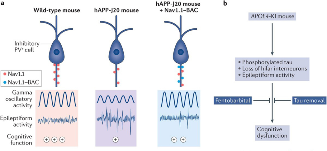
The function of neural circuits and networks can be controlled, in part, by modulating the synchrony of their components' activities. Network hypersynchrony and altered oscillatory rhythmic activity may contribute to cognitive abnormalities in Alzheimer disease (AD). In this condition, network activities that support cognition are altered decades before clinical disease onset, and these alterations predict future pathology and brain atrophy. Although the precise causes and pathophysiological consequences of these network alterations remain to be defined, interneuron dysfunction and network abnormalities have emerged as potential mechanisms of cognitive dysfunction in AD and related disorders. Here, we explore the concept that modulating these mechanisms may help to improve brain function in these conditions.


Figures

Figure 1 |
Figure 1 |. Synchrony and functional states of networks.
The degree of correlated neuronal activity (synchrony) reflects the functional state of networks and circuits. a | To illustrate this relationship at the network level, the top panels show local field potentials (LFPs) recorded from four electrodes (1–4) inserted 1 mm apart into the cat parietal cortex (suprasylvian gyrus) during sleep (left) and wakefulness (right). Network activity during resting and non-active states is predominantly characterized by synchronized slow-frequency and high-amplitude fluctuations (red shading). By contrast, network activity during active states is characterized by desynchronized fast-frequency and low-amplitude fluctuations (blue shading). The bottom panels show hypothetical representations of the amplitude (red line) and frequency (blue line) of LFP fluctuations and of the associated network synchrony (pink line). b | Membrane potential recordings of two layer 2/3 (L2/3) pyramidal neurons from mouse parietal cortex (whisker barrel cortex) during resting and active (whisker use) periods illustrate that neurons desynchronize during active periods. Part a is republished with permission of Society for Neuroscience, from Spatiotemporal analysis of local field potentials and unit discharges in cat cerebral cortex during natural wake and sleep states, Destexhe, A., Contreras, D. & Steriade, M., 19, 11, 1999; permission conveyed through Copyright Clearance Center Inc. Part b is from REF., Nature Publishing Group.
Figure 2 |
Figure 2 |. Encoding reveals network dysfunction in people with mild cognitive impairment.
Memory encoding causes specific changes in brain activity-related functional MRI (fMRI) signals across different networks. People at increased risk for Alzheimer disease (AD) show early changes in the activation and deactivation of specific networks. a | Lateral (left panel) and medial (right panel) views of a healthy left hemibrain. During encoding in healthy people, network activity increases in task-related networks (blue) and decreases in non-task-related networks (yellow/orange). Anti-correlated activity (blue versus yellow/orange) in these two widely distributed and non-overlapping networks is also evident when spontaneous fluctuations of fMRI signals during resting states are examined (not shown). The default mode network (yellow and orange regions) includes brain regions that show decreased fMRI activity during attention-demanding tasks but become active during inwardly oriented mental activity. b | In healthy individuals whose brain activity was monitored by fMRI during a cognitive task, a relatively smaller extent of hippocampal activation and a relatively greater extent of precuneal deactivation were associated with better cognitive performance. c | Pattern separation and completion are cognitive functions that heavily rely on the hippocampal formation and allow us to discriminate (pattern separation) or merge (pattern completion) similar representations or episodes. In a pattern-separation task, in which individuals were asked to discriminate between slightly different trowels (left panel), patients with amnestic mild cognitive impairment (MCI) made more pattern-completion errors (that is, they failed to discriminate slightly different trowels) and had aberrant hyperactivation of the dentate gyrus (DG) and CA3 regions of the hippocampus on fMRI (right panel). Treatment with the antiepileptic drug levetiracetam reversed the hippocampal hyperactivation and improved the patients’ ability to discriminate between images that were similar but not identical. d | During a face–name association task, patients with MCI and amyloid deposits in the brain (as revealed by a Pittsburg compound B-positive (PiB+) signal on positron emission tomography) and patients with AD showed deactivation deficits in the precuneus. Part a is adapted from REF. . Part b is adapted with permission from REF. , Springer. Part c is adapted with permission from REF. , Cell Press/Elsevier. Part d is adapted with permission from REF. , Cell Press/Elsevier.
Figure 3 |
Figure 3 |. Neuronal activity regulates amyloid-β production and deposition.
a | Most patients with Alzheimer disease (AD) and some people without dementia have increased Pittsburg compound B-positive amyloid deposits in the brain (red). Amyloid deposits predominate in brain regions of the default mode network (blue), which shows deactivation deficits in AD (FIG. 2), suggesting a potential link between aberrant neuronal activity and amyloid deposition. b | In APP-A7 mice, chronic, optogenetic stimulation of pyramidal cells in the entorhinal cortex triggered epileptiform activity and increased amyloid deposition in the molecular layer of the dentate gyrus, directly supporting the notion that aberrant neuronal activity can promote amyloid deposition in vivo. c | At the synaptic level, an increase in the frequency of action potentials (APs) proportionally enhances amyloid-β1–42 (Aβ1–42) and amyloid-β1–40 (Aβ1–40) production. Compared with regular firing, burst firing reduces the Aβ1–42/Aβ1–40 ratio. d | Basal neurotransmitter release determines the ‘filter’ mode of synapses and regulates synaptic plasticity. The top panel indicates that excitatory synapses with lower release probability (high-pass-filter synapses) have greater presynaptic Ca2+ build-up, produce lower Aβ1–42/Aβ1–40 ratios, exhibit synaptic facilitation and primarily transfer potentiated responses. The bottom panel depicts synapses with higher release probability (low-pass-filter synapses), which have less presynaptic Ca2+ build-up, produce higher Aβ1–42/Aβ1–40 ratios and show synaptic depression as well as enhanced spike transfer. In AD, synapses may shift from high- to low-pass synaptic filtering. e | Electroencephalograhy recordings capture the combined electrical output of neuronal ensembles. In such recordings, most familial AD (FAD) mice, including hAPP-J20 (REF. 96), APP/PSEN1dE9 (REFS 102,103), Tg2576 (REF. 104), 5xFAD, 3xTg-AD, APP/TTA-EC, APP/TTA-CaMKIIα, and APP23 (REF. 107) mice (Supplementary information S1 (table)), show intermittent large-amplitude epileptiform discharges (denoted by the asterisk in hAPP-J20 mice; bottom panel), which provide evidence of network hypersynchrony. fMRI, functional MRI; PET, positron emission tomography. Part a is republished with permission of Society for Neuroscience, from Molecular, structural, and functional characterization of Alzheimer’s disease: evidence for a relationship between default activity, amyloid, and memory, Buckner, R. L. et al., 25, 34, 2005; permission conveyed through Copyright Clearance Center, Inc. Part e is adapted with permission from REF. , Cell Press/Elsevier.
Figure 4 |
Figure 4 |. Neuronal ensembles generate oscillatory activity patterns.
a | Oscillatory rhythms (grey boxes) emerge from, and control, the activity of neuronal ensembles that are formed by excitatory neurons (blue) and inhibitory interneurons (red). The amplitude, or power, of low-frequency oscillations increases during rest, whereas the amplitude of high-frequency oscillations increases during activity. Action potentials (APs) of excitatory neurons are phase-locked to high-power, lower-frequency oscillations (top grey box), whereas APs of inhibitory cells are phase-locked to high-power, high-frequency oscillations (bottom grey box). b | The left panel shows local field potential (LFP) gamma oscillations (blue) and APs (red) for a parvalbumin-positive (PV+) cell in area CA3, revealing a close association between APs and the phase of gamma oscillations. In the right panel, the AP firing rate (colour coded) is shown as a function of gamma phase for the same PV+ cell, which prominently fires during the ascending phase of gamma oscillations. c | Optogenetic stimulation of PV+ and pyramidal cells at different frequencies resulted in a cell type- and frequency-specific generation of oscillatory activity. 40-Hz stimulation of PV+, but not pyramidal, cells increased gamma power, whereas 8-Hz stimulation of pyramidal, but not PV+, cells increased theta power. d | In macaques, visual stimulation (15s of dim 1 Hz illumination) increased the power of LFP gamma (30–150 Hz) oscillations in a task-related network (visual area V3; left panel) and decreased LFP power across multiple oscillatory frequencies in a default mode network region (posterior cingulate cortex; right panel). SEM, standard error of the mean. Part b is from REF., Nature Publishing Group. Part c is from REF. , Nature Publishing Group. Part d is adapted from Bentley, W. J., Li, J.M., Snyder, A. Z., Raichle, M.E. & Snyder, L.H., Oxygen level and LFP in task-positive and task-negative areas: bridging BOLD fMRI and electrophysiology, Cerebral Cortex, 2014, 26, 1, 346–357, by permission of Oxford Journals.
Figure 5 |
Figure 5 |. Close association between behavioural state, gamma oscillations and epileptiform activity in mice and humans.
a,b | Behavioural activity, full-frequency range spectrograms, gamma oscillatory power, and distribution of epileptic discharges in cortical networks of an hAPP-J20 mouse (part a) and a human with epilepsy (part b). In mice, exploration (active) of a novel environment robustly increased gamma oscillatory power and reduced epileptiform discharges, suggesting that the brain state modulates brain rhythms and network hypersynchrony. In humans, successful, but not unsuccessful, memory encoding also increased gamma oscillatory power and reduced epileptiform discharges. c | In our hypothetical model, memory encoding requires frequency-specific modulation of oscillatory frequencies, which reduces network hypersynchrony. Part a is adapted with permission from REF. , Cell Press/Elsevier. Part b is adapted from Matsumoto, J.Y. et al., Network oscillations modulate interictal epileptiform spike rate during human memory, Brain, 2013, 136, 8, 2444–2456, by permission of Oxford Journals.
Figure 6 |
Figure 6 |. Targeting interneurons to improve Alzheimer disease-related network dysfunction.
a | In hAPP-J20 mice (middle panel), reduced levels of the voltage-gated sodium chanel subunit Nav1.1 in parvalbumin-positive (PV+) cells were associated with reduced gamma power, epileptiform activity and cognitive impairment (the level of cognitive performance is denoted by the plus symbols). Restoring Nav1.1 levels in PV+ cells with an Nav1.1-BAC (bacterial artificial chromosome) transgene (right panel) reduced all of these deficits. b | APOE4-KI mice have increased neuronal levels of phosphorylated tau, age-dependent loss of somatostatin-positive interneurons in the hilus of the dentate gyrus, and seizures. Memory deficits in these mice were reduced by pentobarbital treatment, tau removal and transplantation of interneuron precursor cells. Part a is adapted with permission from REF. , Cell Press/Elsevier.

References

    1. Seidman LJ et al. Medial temporal lobe default mode functioning and hippocampal structure as vulnerability indicators for schizophrenia: A MRI study of non-psychotic adolescent first-degree relatives. Schizophr. Res 159, 426–434 (2014). - PubMed
    1. Fahoum F, Zelmann R, Tyvaert L, Dubeau F. & Gotman J. Epileptic discharges affect the default mode network--fMRI and intracerebral EEG evidence. PLoS One 8, e68038 (2013). - PMC - PubMed
    1. Oser N. et al. Default mode network alterations during language task performance in children with benign epilepsy with centrotemporal spikes (BECTS). Epilepsy Behav. 33, 12–17 (2014). - PubMed
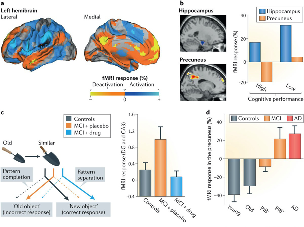
    1. Sperling RA et al. Amyloid deposition is associated with impaired default network function in older persons without dementia. Neuron 63, 178–188 (2009).

      This study shows that PiB+ amyloid deposits are associated with deactivation deficits in a default network component in non-demented individuals and patients with MCI, linking aberrant neuronal activity and pathological changes.

    1. Anticevic A. et al. The role of default network deactivation in cognition and disease. Trends Cogn. Sci 16, 584–592 (2012). - PMC - PubMed