. 2016:2016:6190504.
doi: 10.1155/2016/6190504. Epub 2016 Oct 18.

Activation of ALDH2 with Low Concentration of Ethanol Attenuates Myocardial Ischemia/Reperfusion Injury in Diabetes Rat Model

Affiliations

Activation of ALDH2 with Low Concentration of Ethanol Attenuates Myocardial Ischemia/Reperfusion Injury in Diabetes Rat Model

Pin-Fang Kang et al. Oxid Med Cell Longev. 2016.

Abstract

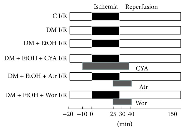
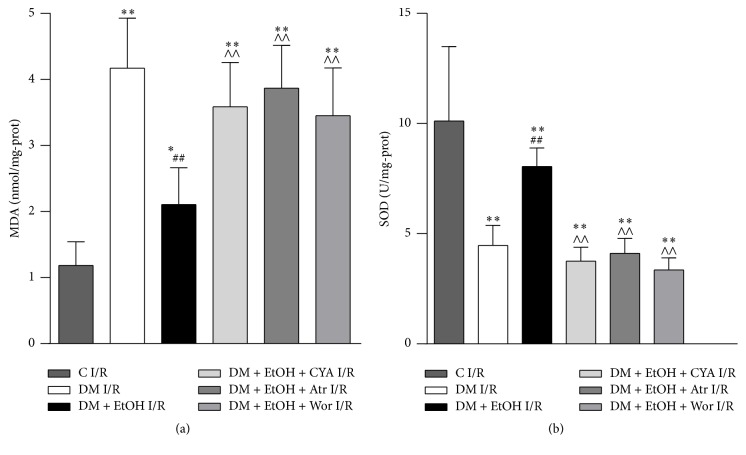
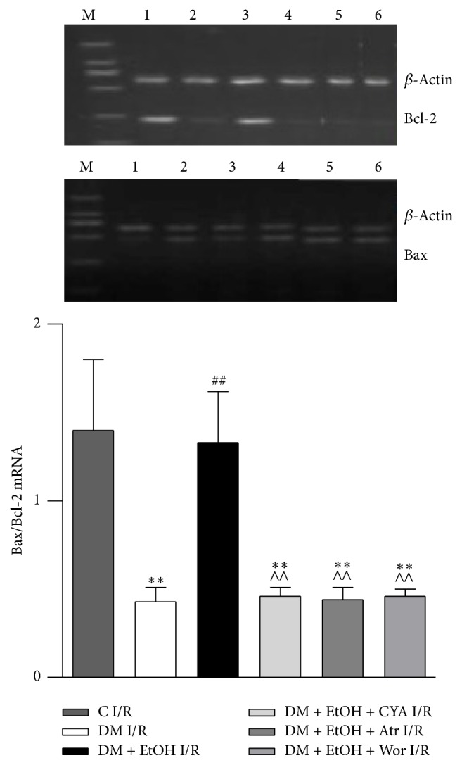
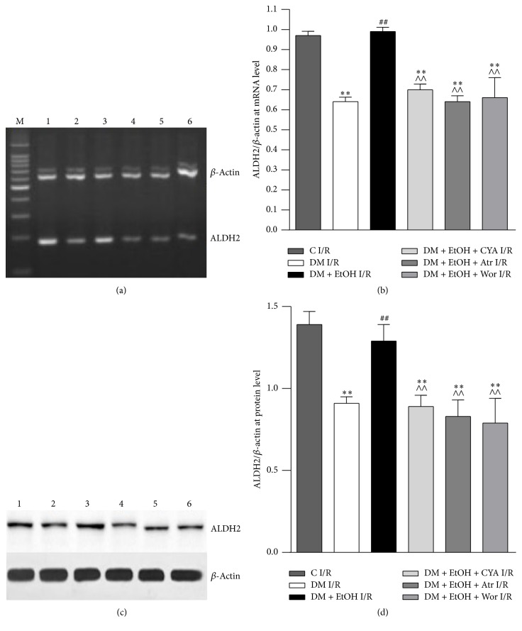
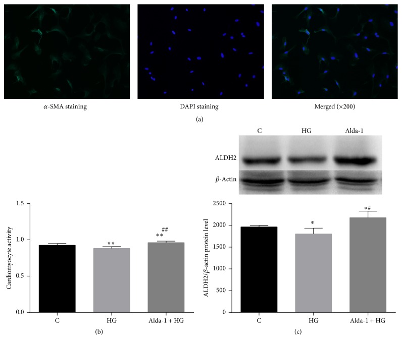
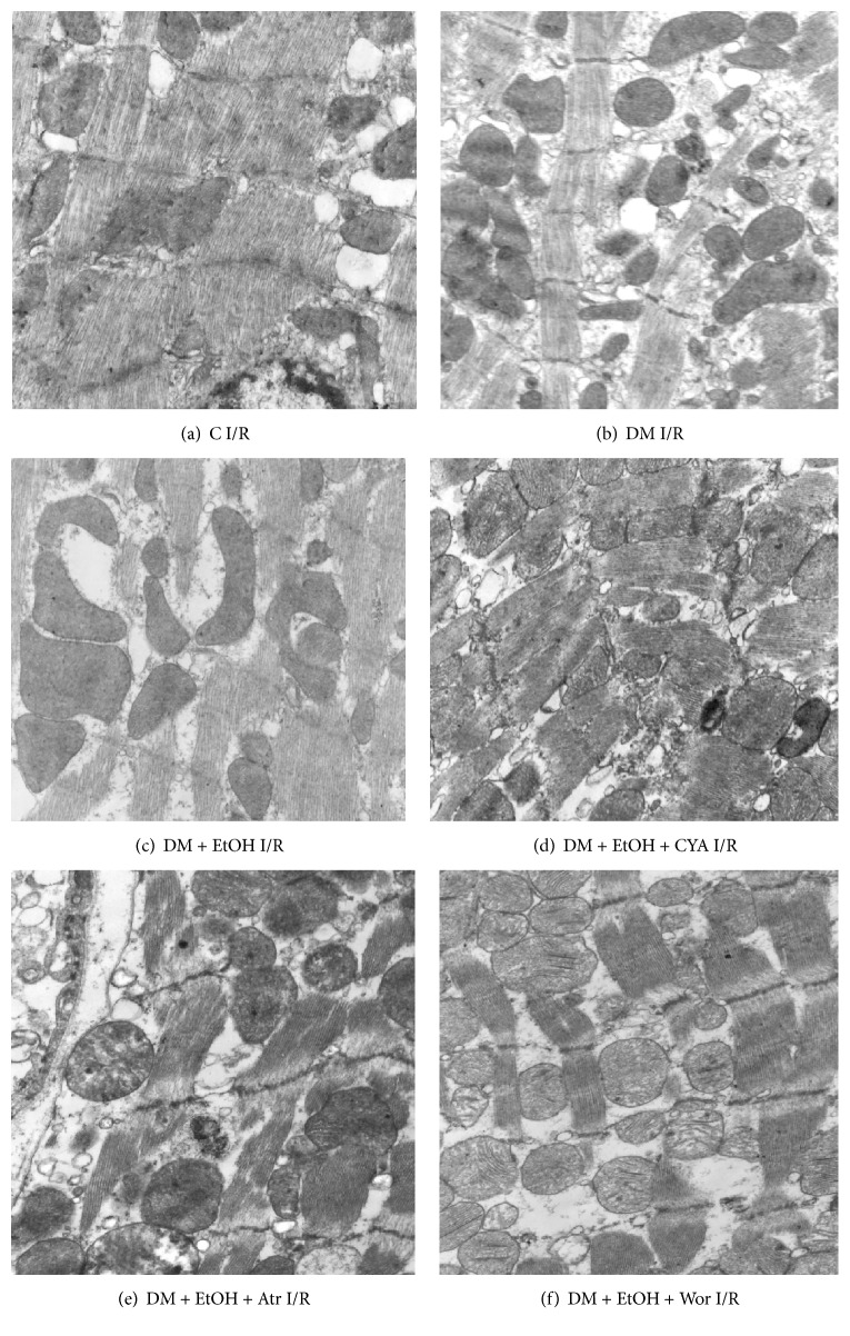
The aim of this paper is to observe the change of mitochondrial aldehyde dehydrogenase 2 (ALDH2) when diabetes mellitus (DM) rat heart was subjected to ischemia/reperfusion (I/R) intervention and analyze its underlying mechanisms. DM rat hearts were subjected to 30 min regional ischemia and 120 min reperfusion in vitro and pretreated with ALDH2 activator ethanol (EtOH); cardiomyocyte in high glucose (HG) condition was pretreated with ALDH2 activator Alda-1. In control I/R group, myocardial tissue structure collapse appeared. Compared with control I/R group, left ventricular parameters, SOD activity, the level of Bcl-2/Bax mRNA, ALDH2 mRNA, and protein expressions were decreased and LDH and MDA contents were increased, meanwhile the aggravation of myocardial structure injury in DM I/R group. When DM I/R rats were pretreated with EtOH, left ventricular parameters, SOD, Bcl-2/Bax, and ALDH2 expression were increased; LDH, MDA, and myocardial structure injury were attenuated. Compared with DM + EtOH I/R group, cyanamide (ALDH2 nonspecific blocker), atractyloside (mitoPTP opener), and wortmannin (PI3K inhibitor) groups all decreased left ventricular parameters, SOD, Bcl-2/Bax, and ALDH2 and increased LDH, MDA, and myocardial injury. When cardiomyocyte was under HG condition, CCK-8 activity and ALDH2 protein expression were decreased. Alda-1 increased CCK-8 and ALDH2. Our findings suggested enhanced ALDH2 expression in diabetic I/R rats played the cardioprotective role, maybe through activating PI3K and inhibiting mitoPTP opening.


Figures

Figure 1
Figure 1
Protocol of various drugs intervention on rat myocardial ischemia/reperfusion model in vitro. C I/R: control ischemia and reperfusion; DM I/R: diabetes rats subjected to myocardial ischemia and reperfusion; DM + EtOH I/R: diabetes + ethanol subjected to myocardial ischemia and reperfusion; DM + EtOH + CYA I/R: diabetes + ethanol + cyanamide subjected to myocardial ischemia and reperfusion; DM + EtOH + Atr I/R: diabetes + ethanol + atractyloside subjected to myocardial ischemia and reperfusion; DM + EtOH + Wor I/R: diabetes + ethanol + wortmannin subjected to myocardial ischemia and reperfusion.
Figure 2
Figure 2
The levels of MDA (a) and SOD (b) of rats myocardium in each group (mean ± SD n = 5). p < 0.05, ∗∗ p < 0.01 versus C I/R; ## p < 0.01 versus DM I/R; ∧∧ p < 0.01 versus DM + EtOH I/R.
Figure 3
Figure 3
The expressions of Bcl-2 and Bax mRNA of heart tissue in rats (mean ± SD of 5 separate experiments). ∗∗ p < 0.01 versus C I/R; ## p < 0.01 versus DM I/R; ∧∧ p < 0.01 versus DM + EtOH I/R. (1) C I/R; (2) DM I/R; (3) DM + EtOH I/R; (4) DM + EtOH + CYA I/R; (5) DM + EtOH + Atr I/R; (6) DM + EtOH + Wor I/R.
Figure 4
Figure 4
Expressions of ALDH2 at mRNA (a, b) and protein (c, d) level of heart tissue in rats (mean ± SD of 5 separate experiments). ∗∗ p < 0.01 versus C I/R; ## p < 0.01 versus DM I/R; ∧∧ p < 0.01 versus DM + EtOH I/R. (1) C I/R; (2) DM I/R; (3) DM + EtOH I/R; (4) DM + EtOH + CYA I/R; (5) DM + EtOH + Atr I/R; (6) DM + EtOH + Wor I/R.
Figure 5
Figure 5
Identify cardiomyocytes by immunofluorescence (a), CCK-8 activity (b), and expressions of ALDH2 protein level of cardiomyocyte in rats (c) (mean ± SD of five separate experiments). p < 0.05, ∗∗ p < 0.01 versus C, # p < 0.05, and ## p < 0.01 versus HG.
Figure 6
Figure 6
Changes of rat myocardium ultrastructural organization in each group (magnification: 10K).

References

    1. Adeghate E., Schattner P., Dunn E. An update on the etiology and epidemiology of diabetes mellitus. Annals of the New York Academy of Sciences. 2006;1084:1–29. doi: 10.1196/annals.1372.029. - DOI - PubMed
    1. Gremizzi C., Vergani A., Paloschi V., Secchi A. Impact of pancreas transplantation on type 1 diabetes-related complications. Current Opinion in Organ Transplantation. 2010;15(1):119–123. doi: 10.1097/MOT.0b013e32833552bc. - DOI - PubMed
    1. Boudina S., Abel E. D. Diabetic cardiomyopathy revisited. Circulation. 2007;115(25):3213–3223. doi: 10.1161/CIRCULATIONAHA.106.679597. - DOI - PubMed
    1. Kooistra M., Geerlings M. I., Mali W. P. T. M., Vincken K. L., Van Der Graaf Y., Biessels G. J. Diabetes mellitus and progression of vascular brain lesions and brain atrophy in patients with symptomatic atherosclerotic disease. The SMART-MR study. Journal of the Neurological Sciences. 2013;332(1-2):69–74. doi: 10.1016/j.jns.2013.06.019. - DOI - PubMed
    1. Zhang Y., Ren J. ALDH2 in alcoholic heart diseases: molecular mechanism and clinical implications. Pharmacology and Therapeutics. 2011;132(1):86–95. doi: 10.1016/j.pharmthera.2011.05.008. - DOI - PMC - PubMed

MeSH terms

LinkOut - more resources