. 2016 Oct 15;8(10):4195-4204.
eCollection 2016.

Effect of bisphenol a on occurrence and progression of prolactinoma and its underlying mechanisms

Affiliations

Effect of bisphenol a on occurrence and progression of prolactinoma and its underlying mechanisms

Lanxiang Hao et al. Am J Transl Res. .

Abstract

Objective: To investigate the effects of Bisphenol A (BPA) on prolactin (PRL) release, pituitary cell proliferation, prolactinoma formation in estrogen-sensitive Fischer 344 (F344) rats.

Materials and methods: Four-week-old female F344 rats were orally administered with different concentrations of BPA or intraperitoneal injection of estradiol benzoate (estradiolbenzoate, E2) for 12 weeks. Bodyweight, blood RPL level and pituitary weights were observed and recorded. Real-time PCR, western blot and immunohistochemistry analysis were used to detect the mRNA and protein levels of the proliferation markers, including proliferating cell neclear antigen (PCNA), pituitary tumor-transforming gene (PTTG) and its relevant marker ERα. Plasma and urine BPA concentration in patients with prolactinoma and healthy participants were measured as well.

Results: Body weights of the rats treated with BPA were significantly decreased compared with those in the control group. The plasma PRL level and the pituitary weights of the rats were higher than those in the control group after BPA treatment. Compared with the control group, the pituitary mRNA and protein expression levels of PCNA and PTTG were significantly increased after BPA treatment. Moreover, ERα expression level was enhanced by the treatment of BPA in comparison with that of the control group. Finally, the plasma BPA concentration in the prolactin tumor patients was significantly higher than that in the healthy participants.

Conclusion: BPA can significantly promote pituitary cell proliferation and prolactin secretion in F344 rats, which may have impact on the proliferation and secretion of pituitary cell function through the ERα pathway.

Keywords: Prolactinomas; bisphenol A; cell proliferation; estradiol; prolactin secretion.


Figures

Figure 1
Figure 1
BPA exposure increased pituitary weight plasma PRL level in rat. A. The pituitary weight changes after rat treat with BPA 50 mg/kg/d, BPA 200 mg/kg/d, BPA 400 mg/kg/d and E2. B. The PRL level in the serum of the rats from indicated groups with different treatment 6 weeks. C. The PRL level in the serum of the rats with indicated treatment. Experiment for each sample was repeated for at least three times. Significant differences between control group experimental groups are shown by “*” to indicate significant difference (P<0.05).
Figure 2
Figure 2
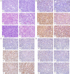
BPA and E2 exposure promoted the formation of pituitary tumor. A. The HE staining of rat pituitary gland Indicated that BPA and E2 could promoted the formation of pituitary tumor; B. The immunohistochemical staining of pituitary PRL level; C. The immunohistochemical staining for pituitary PCNA; D. The immunohistochemical staining for pituitary ERα. Group set: A (Control), B (Oil control), C (BPA50), D (BPA200), E (BPA400), F (E2).
Figure 3
Figure 3
The western blot results of rat pituitary PTTG, PCNA, ERα, ERβ. A: Western blot analysis for PTTG and ERα; B: Western blot analysis for PCNA and ERβ.
Figure 4
Figure 4
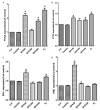
Quantification analysis of the WB blot data for different genes. A: Quantification analysis of rat pituitary PCNA. B. Quantification analysis of rat pituitary PTTG. C: Quantification analysis of rat pituitary ERα. D: Quantification analysis of rat pituitary ERβ.
Figure 5
Figure 5
qPCR analysis for mRNA expression of indicated genes among different groups. A. The results of PCNA mRNA expression in pituitaries; B. The results of PTTG mRNA expression in pituitaries; C. The results of ERα mRNA expression in pituitaries; D. The results of ERβ mRNA expression in pituitaries. Experiment for each sample was repeated for at least three times. Significant differences between control group experimental groups are shown by “*” to indicate significant difference (P<0.05).
Figure 6
Figure 6
Serum BPA concentration was significant evaluated in prolactinoma patients. A. The BPA concentration in serum of the prolactinoma patients and healthy controls; B. The BPA concentration in urine from the prolactinoma patients and healthy control; Experiment for each sample was repeated for at least three times. Significant differences between control group experimental groups are shown by “*” to indicate significant difference (P<0.05).

References

    1. Glezer A, Bronstein MD. Prolactinomas. Endocrinol Metab Clin North Am. 2015;44:71–78. - PubMed
    1. Gillam MP, Molitch ME, Lombardi G, Colao A. Advances in the treatment of prolactinomas. Endocr Rev. 2006;27:485–534. - PubMed
    1. Schlechte JA. Clinical practice. Prolactinoma. N Engl J Med. 2003;349:2035–2041. - PubMed
    1. Takekoshi S, Yasui Y, Inomoto C, Kitatani K, Nakamura N, Osamura RY. A Histopathological Study of Multi-hormone Producing Proliferative Lesions in Estrogen-induced Rat Pituitary Prolactinoma. Acta Histochem Cytochem. 2014;47:155–164. - PMC - PubMed
    1. Cao L, Gao H, Gui S, Bai G, Lu R, Wang F, Zhang Y. Effects of the estrogen receptor antagonist fulvestrant on F344 rat prolactinoma models. J Neurooncol. 2014;116:523–531. - PubMed

LinkOut - more resources