

Cellular mechanical properties reflect the differentiation potential of nucleus pulposus-derived progenitor cells
- PMID: 27830029
- PMCID: PMC5095338
Cellular mechanical properties reflect the differentiation potential of nucleus pulposus-derived progenitor cells
Abstract
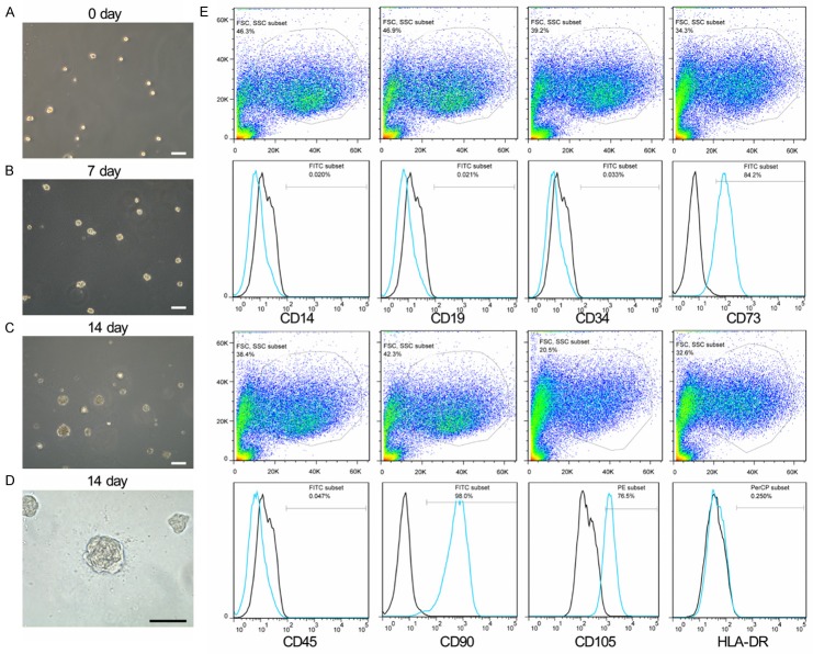
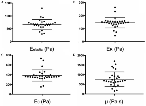
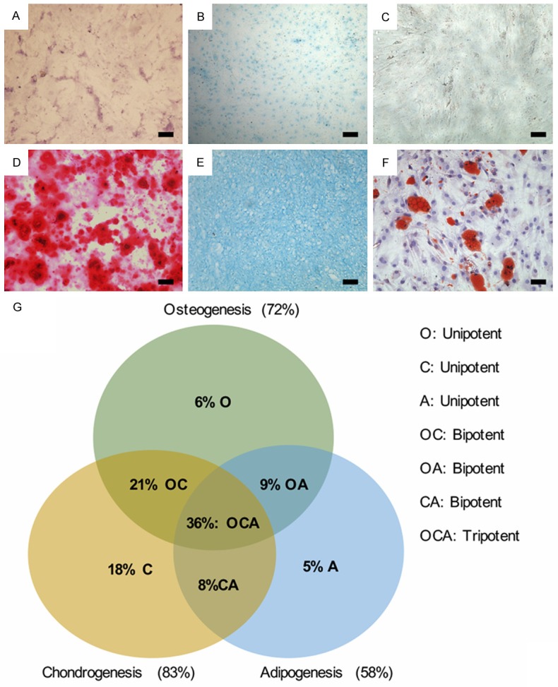
Mechanical properties of cells reflect differences in cellular subpopulations, differentiation potency, and cell behaviors. Previous study has revealed that intervertebral disc (IVD) degeneration leads to alterations in cell behavior and differentiation potency. Human nucleus pulposus-derived progenitor cells (NPPCs) are an attractive cell sources for IVD regeneration. However, the relationship between mechanical properties and differentiation potential in different NPPC subpopulations is few known. In this study, mechanical properties of different NPPC subpopulations were measured via atomic force microscopy (AFM) and correlated with differentiation potential of NPPCs. We found that elastic modulus, relaxed modulus, and instantaneous modulus were positively correlated with osteogenic potential of NPPCs. And apparent viscosity was correlated with chondrogenic potential of NPPCs. These results indicated that the mechanical properties were predictive markers for differentiation potential of NPPC subpopulations, and could be used for enrichment based on differentiation potential, which could significantly improve the outcome of IVD regeneration.
Keywords: Cellular mechanical properties; intervertebral disc degeneration; nucleus pulposus; stem cell.
Figures
Similar articles
-
Intervertebral disc-derived stem cells: implications for regenerative medicine and neural repair.Spine (Phila Pa 1976). 2013 Feb 1;38(3):211-6. doi: 10.1097/BRS.0b013e318266a80d. Spine (Phila Pa 1976). 2013. PMID: 22772571
-
Differentiation of Pluripotent Stem Cells into Nucleus Pulposus Progenitor Cells for Intervertebral Disc Regeneration.Curr Stem Cell Res Ther. 2019;14(1):57-64. doi: 10.2174/1574888X13666180918095121. Curr Stem Cell Res Ther. 2019. PMID: 30227822 Review.
-
Rejuvenation Modulation of Nucleus Pulposus Progenitor Cells Reverses Senescence-Associated Intervertebral Disc Degeneration.Adv Mater. 2025 Feb;37(7):e2409979. doi: 10.1002/adma.202409979. Epub 2024 Dec 29. Adv Mater. 2025. PMID: 39969420
-
Characterization of the Nucleus Pulposus Progenitor Cells via Spatial Transcriptomics.Adv Sci (Weinh). 2024 May;11(18):e2303752. doi: 10.1002/advs.202303752. Epub 2024 Feb 4. Adv Sci (Weinh). 2024. PMID: 38311573 Free PMC article.
-
Mechanisms of endogenous repair failure during intervertebral disc degeneration.Osteoarthritis Cartilage. 2019 Jan;27(1):41-48. doi: 10.1016/j.joca.2018.08.021. Epub 2018 Sep 19. Osteoarthritis Cartilage. 2019. PMID: 30243946 Review.
Cited by
-
Autophagy mediates serum starvation-induced quiescence in nucleus pulposus stem cells by the regulation of P27.Stem Cell Res Ther. 2019 Apr 15;10(1):118. doi: 10.1186/s13287-019-1219-8. Stem Cell Res Ther. 2019. PMID: 30987681 Free PMC article.
-
IVD progenitor cells: a new horizon for understanding disc homeostasis and repair.Nat Rev Rheumatol. 2019 Feb;15(2):102-112. doi: 10.1038/s41584-018-0154-x. Nat Rev Rheumatol. 2019. PMID: 30643232 Review.
References
-
- Luoma K, Riihimaki H, Luukkonen R, Raininko R, Viikari-Juntura E, Lamminen A. Low back pain in relation to lumbar disc degeneration. Spine (Phila Pa 1976) 2000;25:487–492. - PubMed
-
- Takatalo J, Karppinen J, Niinimaki J, Taimela S, Nayha S, Mutanen P, Sequeiros RB, Kyllonen E, Tervonen O. Does lumbar disc degeneration on magnetic resonance imaging associate with low back symptom severity in young Finnish adults? Spine (Phila Pa 1976) 2011;36:2180–2189. - PubMed
-
- Sakai D, Andersson GB. Stem cell therapy for intervertebral disc regeneration: obstacles and solutions. Nat Rev Rheumatol. 2015;11:243–256. - PubMed
-
- Blanco JF, Graciani IF, Sanchez-Guijo FM, Muntion S, Hernandez-Campo P, Santamaria C, Carrancio S, Barbado MV, Cruz G, Gutierrez-Cosio S, Herrero C, San Miguel JF, Brinon JG, del Canizo MC. Isolation and characterization of mesenchymal stromal cells from human degenerated nucleus pulposus: comparison with bone marrow mesenchymal stromal cells from the same subjects. Spine (Phila Pa 1976) 2010;35:2259–2265. - PubMed
LinkOut - more resources
Full Text Sources
Miscellaneous