

Human immunodeficiency virus (HIV) is highly associated with giant idiopathic esophageal ulcers in acquired immunodeficiency syndrome (AIDS) patients
- PMID: 27830031
- PMCID: PMC5095340
Human immunodeficiency virus (HIV) is highly associated with giant idiopathic esophageal ulcers in acquired immunodeficiency syndrome (AIDS) patients
Abstract
Objective: This study aimed to determine whether the human immunodeficiency virus (HIV) exists in giant idiopathic esophageal ulcers in the patients with acquired immune deficiency syndrome (AIDS).
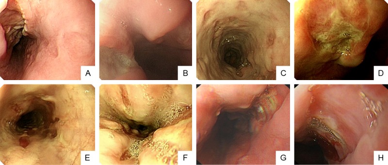
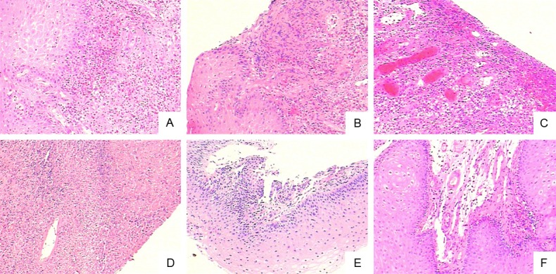
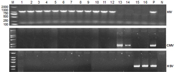
Methods: 16 AIDS patients with a primary complaint of epigastric discomfort were examined by gastroscopy. Multiple and giant esophageal ulcers were biopsied and analyzed with pathology staining and reverse transcription-polymerase chain reaction (RT-PCR) to determine the potential pathogenic microorganisms, including HIV, cytomegalovirus (CMV) and herpes simplex viruses (HSV).
Results: HIV was detected in ulcer samples from 12 out of these 16 patients. Ulcers in 2 patients were infected with CMV and ulcers in another 2 patients were found HSV positive. No obvious cancerous pathological changes were found in these multiple giant esophageal ulcer specimens.
Conclusion: HIV may be one of the major causative agents of multiple benign giant esophageal ulcers in AIDS patients.
Keywords: AIDS; CMV; HIV; HSV; endoscopy; esophageal ulcer.
Figures
Similar articles
-
Advantages and pitfalls of the polymerase chain reaction in the diagnosis of esophageal ulcers in AIDS patients.Dig Dis Sci. 2009 Sep;54(9):1933-9. doi: 10.1007/s10620-008-0584-4. Epub 2008 Dec 3. Dig Dis Sci. 2009. PMID: 19051024 Clinical Trial.
-
[Esophageal pathology in patients with the AIDS virus. Etiology and diagnosis].Acta Gastroenterol Latinoam. 1991;21(2):67-83. Acta Gastroenterol Latinoam. 1991. PMID: 1820692 Spanish.
-
Diagnosis of esophageal ulcers in acquired immunodeficiency syndrome.Semin Gastrointest Dis. 1999 Jul;10(3):85-92. Semin Gastrointest Dis. 1999. PMID: 10435695
-
HIV-1 gp41 antigen demonstration in esophageal ulcers with acquired immunodeficiency syndrome.J Clin Gastroenterol. 1991 Dec;13(6):644-8. doi: 10.1097/00004836-199112000-00007. J Clin Gastroenterol. 1991. PMID: 1761837 Review.
-
Multi-step pathogenesis of AIDS--role of cytomegalovirus.Res Immunol. 1991 Feb;142(2):87-95. doi: 10.1016/0923-2494(91)90016-c. Res Immunol. 1991. PMID: 1650955 Review.
Cited by
-
Upper Gastrointestinal Bleeding Due to Idiopathic Oesophageal Ulceration in the Era of HAART: A Vanishing yet Pernicious Aetiology.J Clin Diagn Res. 2017 May;11(5):OD20-OD21. doi: 10.7860/JCDR/2017/26205.9902. Epub 2017 May 1. J Clin Diagn Res. 2017. PMID: 28658836 Free PMC article.
-
The spectrum of esophagitis in patients living with HIV - a scoping review.Germs. 2024 Jun 30;14(2):188-196. doi: 10.18683/germs.2024.1430. eCollection 2024 Jun. Germs. 2024. PMID: 39493738 Free PMC article.
References
-
- Wilcox CM, Rodgers W, Lazenby A. Prospective comparison of brush cytology, viral culture, and histology for the diagnosis of ulcerative esophagitis in AIDS. Clin Gastroenterol Hepatol. 2004;2:564–567. - PubMed
-
- Dragean CA, Bogdan I, Azzouzzi K, Goncette L. Giant idiopathic ulcer of esophagus in the context of acquired immunodeficiency syndrome (AIDS) JBR-BTR. 2013;96:72–74. - PubMed
-
- Brunaldi MO, Rezende RE, Garcia SB, Machado AA, Modena JL, Zucoloto S. Esophageal ulcer in Brazilian patients with HIV: prevalence and comparative analysis among diagnostic methods. AIDS Patient Care STDS. 2010;24:311–316. - PubMed
-
- Wang LW, Ma YL, Lv BL, Xu YH, Zhou MZ, Qiu CL, Huang SP, Shen YZ, Zhang RF, Cheng JL. One case with multiple huge esophageal ulcers caused by HIV infection and literature review. Chinese Journal of Digestive Endoscopy. 2012:166–167.
-
- Victoria JM, Guimaraes AL, da Silva LM, Kalapothakis E, Gomez RS. Polymerase chain reaction for identification of herpes simplex virus (HSV-1), cytomegalovirus (CMV) and human herpes virus-type 6 (HHV-6) in oral swabs. Microbiol Res. 2005;160:61–65. - PubMed
LinkOut - more resources
Full Text Sources