. 2016:2016:5742346.
doi: 10.1155/2016/5742346. Epub 2016 Oct 17.

Active Pedicle Epithelial Flap Transposition Combined with Amniotic Membrane Transplantation for Treatment of Nonhealing Corneal Ulcers

Affiliations

Active Pedicle Epithelial Flap Transposition Combined with Amniotic Membrane Transplantation for Treatment of Nonhealing Corneal Ulcers

Ting Zhang et al. J Ophthalmol. 2016.

Abstract

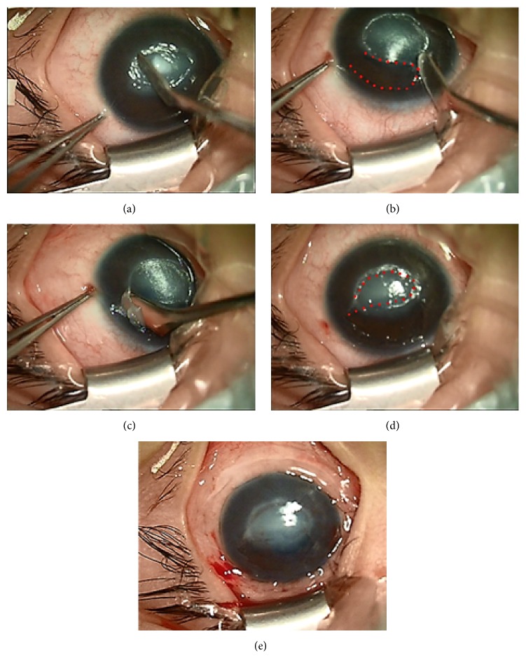
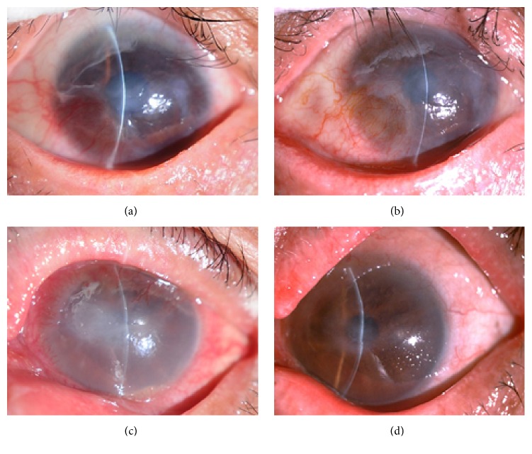
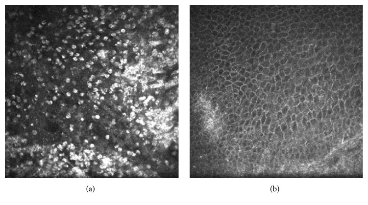
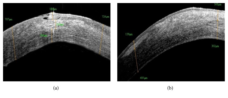
Introduction. The objective was to evaluate the efficacy of active pedicle epithelial flap transposition combined with amniotic membrane transplantation (AMT) in treating nonhealing corneal ulcers. Material and Methods. Eleven patients (11 eyes) with nonhealing corneal ulcer who underwent the combined surgery were included. Postoperatively, ulcer healing time was detected by corneal fluorescein staining. Visual acuity, intraocular pressure, surgical complications, and recurrence were recorded. Corneal status was inspected by the laser scanning confocal microscopy and anterior segment optical coherence tomography (AS-OCT). Results. The primary diseases were herpes simplex keratitis (8 eyes), corneal graft ulcer (2 eyes), and Stevens-Johnson syndrome (1 eye). All epithelial flaps were intact following surgery, without shedding or displacement. Mean ulcer healing time was 10.8 ± 3.1 days, with a healing rate of 91%. Vision significantly improved from 1.70 to 0.82 log MAR (P = 0.001). A significant decrease in inflammatory cell infiltration and corneal stromal edema was revealed 2 months postoperatively by confocal microscopy and AS-OCT. Corneal ulcer recurred in 1 eye. None of the patients developed major complications. Conclusion. Active pedicle epithelial flap transposition combined with AMT is a simple and effective treatment for nonhealing corneal ulcers.


Figures

Figure 1
Figure 1
Photographs showing the procedures of epithelial flap transposition combined with amniotic membrane transplantation. (a) Cleaning the ulcer surface. (b) Constructing a flap boundary (the dotted line demonstrates the region from where the epithelial flap was obtained). (c) Obtaining an epithelial flap. (d) Transposing the epithelial flap (the dotted line demonstrates the translocated epithelial flap). (e) Suturing an amniotic membrane patch.
Figure 2
Figure 2
Slit-lamp photographs showing the treatment course of nonhealing corneal ulcers. (a) Patient 1: the preoperative uncorrected visual acuity was finger counting. The size of the ulcer was approximately 4.0 × 3.0 mm. The surrounding tissue displayed infiltration and edema. (b) At 1 month after surgery, the corneal ulcer was completely cured, with the relief of corneal opacity, and the postoperative uncorrected visual acuity was 0.1 (1.0 log MAR). (c) Patient 2: the preoperative uncorrected visual acuity was hand motion. The ulcer was approximately 5.0 × 2.5 mm. Evident stromal necrosis, edema, and anterior chamber hypopyon were observed. (d) At 1 month after surgery, the corneal ulcer was healed, with the relief of corneal edema, and the postoperative uncorrected visual acuity was 0.1 (1.0 log MAR).
Figure 3
Figure 3
(a) Abundant inflammatory cells were displayed in the epithelium and basal membrane surrounding the epithelial nonhealing region of the ulcer. (b) The number of inflammatory cells decreased significantly at 2 months after surgery.
Figure 4
Figure 4
(a) The ulcer was shown to involve approximately 1/3 of the corneal stroma, with stromal edema and infiltration. (b) The combined surgery healed the ulcer, leaving a semitransparent and highly reflective region in the cornea at 2 months postoperatively.

Similar articles

Cited by

References

    1. Mohan S., Budhiraja I., Saxena A., Khan P., Sachan S. K. Role of multilayered amniotic membrane transplantation for the treatment of resistant corneal ulcers in North India. International Ophthalmology. 2014;34(3):485–491. doi: 10.1007/s10792-013-9834-3. - DOI - PubMed
    1. Livingstone I., Stefanowicz F., Moggach S., et al. New insight into non-healing corneal ulcers: iatrogenic crystals. Eye. 2013;27(6):755–762. doi: 10.1038/eye.2013.39. - DOI - PMC - PubMed
    1. Prabhasawat P., Tesavibul N., Komolsuradej W. Single and multilayer amniotic membrane transplantation for persistent corneal epithelial defect with and without stromal thinning and perforation. British Journal of Ophthalmology. 2001;85(12):1455–1463. doi: 10.1136/bjo.85.12.1455. - DOI - PMC - PubMed
    1. Gabison E. E., Doan S., Catanese M., Chastang P., Ben M'hamed M., Cochereau I. Modified deep anterior lamellar keratoplasty in the management of small and large epithelialized descemetoceles. Cornea. 2011;30(10):1179–1182. doi: 10.1097/ICO.0b013e3182031c81. - DOI - PubMed
    1. Medsinge A., Gajdosova E., Moore W., Nischal K. K. Management of inflammatory corneal melt leading to central perforation in children: a retrospective study and review of literature. Eye. 2016;30(4):593–601. doi: 10.1038/eye.2015.278. - DOI - PMC - PubMed

LinkOut - more resources