

Reliability of measuring regional callosal atrophy in neurodegenerative diseases
- PMID: 27830115
- PMCID: PMC5094205
- DOI: 10.1016/j.nicl.2016.10.012
Reliability of measuring regional callosal atrophy in neurodegenerative diseases
Abstract
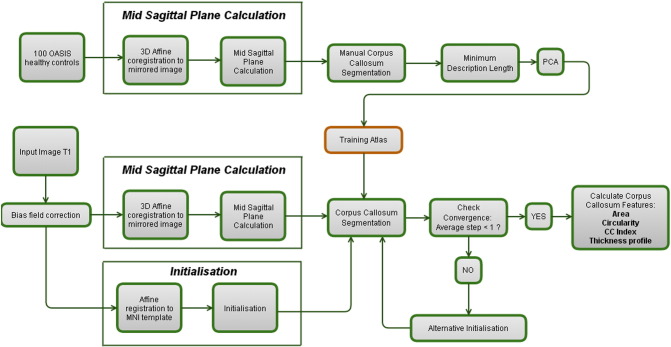
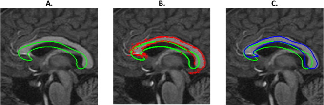
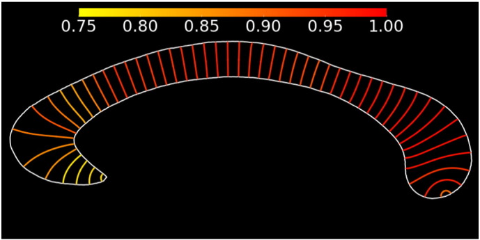
The Corpus Callosum (CC) is an important structure connecting the two brain hemispheres. As several neurodegenerative diseases are known to alter its shape, it is an interesting structure to assess as biomarker. Yet, currently, the CC-segmentation is often performed manually and is consequently an error prone and time-demanding procedure. In this paper, we present an accurate and automated method for corpus callosum segmentation based on T1-weighted MRI images. After the initial construction of a CC atlas based on healthy controls, a new image is subjected to a mid-sagittal plane (MSP) detection algorithm and a 3D affine registration in order to initialise the CC within the extracted MSP. Next, an active shape model is run to extract the CC. We calculated the reliability of most popular CC features (area, circularity, corpus callosum index and thickness profile) in healthy controls, Alzheimer's Disease patients and Multiple Sclerosis patients. Importantly, we also provide inter-scanner reliability estimates. We obtained an intra-class correlation coefficient (ICC) of over 0.95 for most features and most datasets. The inter-scanner reliability assessed on the MS patients was remarkably well and ranged from 0.77 to 0.97. In summary, we have constructed an algorithm that reliably detects the CC in 3D T1 images in a fully automated way in healthy controls and different neurodegenerative diseases. Although the CC area and the circularity are the most reliable features (ICC > 0.97); the reliability of the thickness profile (ICC > 0.90; excluding the tip) is sufficient to warrant its inclusion in future clinical studies.
Keywords: Alzheimer's disease; Biomarker; Corpus callosum segmentation; Corpus callosum thickness profile; Multiple sclerosis; Repeatability; Reproducibility.
Figures
References
-
- Aboitiz F. Brain connections: interhemispheric fiber systems and anatomical brain asymmetries in humans. Biol. Res. 1992;25:51–61. - PubMed
-
- Adamson C., Beare R., Walterfang M., Seal M. Software pipeline for midsagittal corpus callosum thickness profile processing: automated segmentation, manual editor, thickness profile generator, group-wise statistical comparison and results display. Neuroinformatics. 2014;12:595–614. - PubMed
-
- Ardekani B.A., Guckemus S., Bachman A., Hoptman M.J., Wojtaszek M., Nierenberg J. Quantitative comparison of algorithms for inter-subject registration of 3D volumetric brain MRI scans. J. Neurosci. Methods. 2005;142:67–76. - PubMed
Publication types
MeSH terms
Substances
Grants and funding
LinkOut - more resources
Full Text Sources
Other Literature Sources
Medical
Research Materials
