

Qualitative evaluation of anterior segment in angle closure disease using anterior segment optical coherence tomography
- PMID: 27830199
- PMCID: PMC5093787
- DOI: 10.1016/j.joco.2016.06.005
Qualitative evaluation of anterior segment in angle closure disease using anterior segment optical coherence tomography
Abstract
Purpose: To evaluate different mechanisms of primary angle closure (PAC) and to quantify anterior chamber (AC) parameters in different subtypes of angle closure disease using anterior segment optical coherence tomography (AS-OCT).
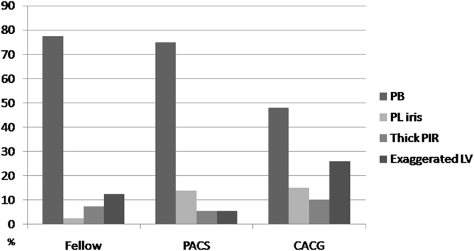
Methods: In this prospective study, 115 eyes of 115 patients with angle closure disease were included and categorized into three groups: 1) fellow eyes of acute angle closure (AAC; 40 eyes); 2) primary angle closure glaucoma (PACG; 39 eyes); and 3) primary angle closure suspect (PACS; 36 eyes). Complete ophthalmic examination including gonioscopy, A-scan biometry, and AS-OCT were performed. Based on the AS-OCT images, 4 mechanisms of PAC including pupillary block, plateau iris configuration, thick peripheral iris roll (PIR), and exaggerated lens vault were evaluated. Angle, AC, and lens parameter variables were also evaluated among the three subtypes.
Results: There was a statistically significant difference in the mechanism of angle closure among the three groups (p = 0.03). While the majority of fellow eyes of AAC and of PACS eyes had pupillary block mechanism (77.5% and 75%, respectively), only 48.7% of PACG eyes had dominant pupillary block mechanism (p = 0.03). The percentage of exaggerated lens vault and plateau iris mechanisms was higher in PACG eyes (25.5% and 15.4%, respectively). Fellow eyes of AAC had the shallowest AC (p = 0.01), greater iris curvature (p = 0.01), and lens vault (p = 0.02) than PACS and PACG eyes. Iris thickness was not significantly different among the three groups (p = 0.45).
Conclusion: Using AS-OCT, we found that there was a statistically significant difference in the underlying PAC mechanisms and quantitative AC parameters among the three subtypes of angle closure disease.
Keywords: Angle closure; Anterior segment optical coherence tomography; Glaucoma; Iris curvature; Lens vault.
Figures
Similar articles
-
Acute angle closure: qualitative and quantitative evaluation of the anterior segment using anterior segment optical coherence tomography.Clin Exp Ophthalmol. 2014 Sep-Oct;42(7):615-22. doi: 10.1111/ceo.12285. Epub 2014 Jan 27. Clin Exp Ophthalmol. 2014. PMID: 24330237
-
Role of lens vault in subtypes of angle closure in Iranian subjects.Eye (Lond). 2014 Mar;28(3):337-43. doi: 10.1038/eye.2013.296. Epub 2014 Jan 10. Eye (Lond). 2014. PMID: 24406416 Free PMC article.
-
Ocular biometry in the subtypes of angle closure: an anterior segment optical coherence tomography study.Am J Ophthalmol. 2013 Apr;155(4):664-673, 673.e1. doi: 10.1016/j.ajo.2012.10.014. Epub 2012 Dec 13. Am J Ophthalmol. 2013. PMID: 23246271
-
Anterior Segment Optical Coherence Tomography Changes to the Anterior Chamber Angle in the Short-term following Laser Peripheral Iridoplasty.J Curr Glaucoma Pract. 2014 Jan-Apr;8(1):1-6. doi: 10.5005/jp-journals-10008-1152. Epub 2014 Jan 16. J Curr Glaucoma Pract. 2014. PMID: 26997799 Free PMC article. Review.
-
Primary angle closure glaucoma in Chinese and Western populations.Chin Med J (Engl). 2002 Nov;115(11):1706-15. Chin Med J (Engl). 2002. PMID: 12609093 Review.
Cited by
-
Lens-vault analysis and its correlation with other biometric parameters using swept-source OCT.J Optom. 2022 Jan-Mar;15(1):88-99. doi: 10.1016/j.optom.2021.04.001. Epub 2021 Nov 1. J Optom. 2022. PMID: 34736867 Free PMC article.
-
Automated classification of angle-closure mechanisms based on anterior segment optical coherence tomography images via deep learning.Heliyon. 2024 Jul 26;10(15):e35236. doi: 10.1016/j.heliyon.2024.e35236. eCollection 2024 Aug 15. Heliyon. 2024. PMID: 39166052 Free PMC article.
-
One-year outcomes of combined phacoemulsification and viscogoniosynechialysis with and without endoscopic cyclophotocoagulation in primary angle-closure glaucoma.Int Ophthalmol. 2023 Sep;43(9):3227-3236. doi: 10.1007/s10792-023-02723-0. Epub 2023 Apr 18. Int Ophthalmol. 2023. PMID: 37071345
-
Diurnal Variation of Optical Coherence Tomography Measurements of Static and Dynamic Anterior Segment Parameters.J Glaucoma. 2018 Jan;27(1):16-21. doi: 10.1097/IJG.0000000000000832. J Glaucoma. 2018. PMID: 29194197 Free PMC article.
-
Differences in Anterior Chamber Angle Assessments Between Gonioscopy, EyeCam, and Anterior Segment OCT: The Chinese American Eye Study.Transl Vis Sci Technol. 2019 Mar 26;8(2):5. doi: 10.1167/tvst.8.2.5. eCollection 2019 Mar. Transl Vis Sci Technol. 2019. PMID: 30941263 Free PMC article.
References
-
- Niwas S.I., Lin W., Bai X. Reliable feature selection for automated angle closure glaucoma mechanism detection. J Med Syst. 2015;39:1–10. - PubMed
-
- Shabana N., Aquino M.C.D., See J. Quantitative evaluation of anterior chamber parameters using anterior segment optical coherence tomography in primary angle closure mechanisms. Clin Exp Ophthalmol. 2012;40:792–801. - PubMed
-
- Sng C.C., Aquino M.C.D., Liao J. Pretreatment anterior segment imaging during acute primary angle closure: insights into angle closure mechanisms in the acute phase. Ophthalmology. 2014;121:119–125. - PubMed
-
- Tarongoy P., Ho C.L., Walton D.S. Angle-closure glaucoma: the role of the lens in the pathogenesis, prevention, and treatment. Surv Ophthalmol. 2009;54:211–225. - PubMed
LinkOut - more resources
Full Text Sources
Other Literature Sources
Research Materials
