

Separating melanin from hemodynamics in nevi using multimode hyperspectral dermoscopy and spatial frequency domain spectroscopy
- PMID: 27830262
- PMCID: PMC5103103
- DOI: 10.1117/1.JBO.21.11.114001
Separating melanin from hemodynamics in nevi using multimode hyperspectral dermoscopy and spatial frequency domain spectroscopy
Abstract
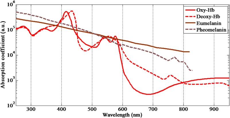
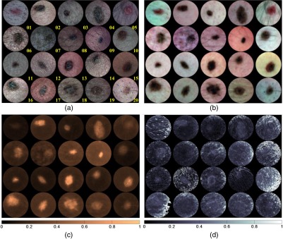
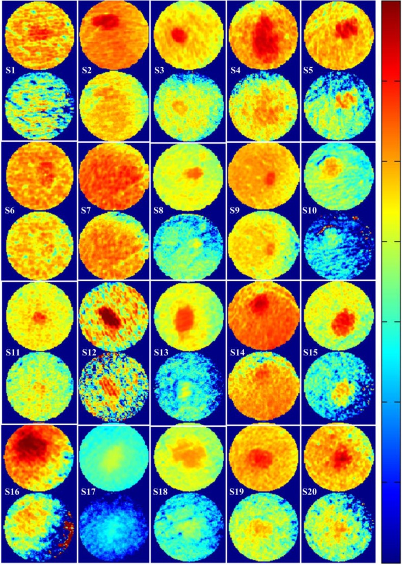
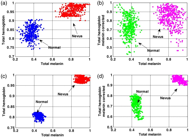
Changes in the pattern and distribution of both melanocytes (pigment producing) and vasculature (hemoglobin containing) are important in distinguishing melanocytic proliferations. The ability to accurately measure melanin distribution at different depths and to distinguish it from hemoglobin is clearly important when assessing pigmented lesions (benign versus malignant). We have developed a multimode hyperspectral dermoscope (SkinSpect™) able to more accurately image both melanin and hemoglobin distribution in skin. SkinSpect uses both hyperspectral and polarization-sensitive measurements. SkinSpect’s higher accuracy has been obtained by correcting for the effect of melanin absorption on hemoglobin absorption in measurements of melanocytic nevi. In vivo human skin pigmented nevi (N=20) were evaluated with the SkinSpect, and measured melanin and hemoglobin concentrations were compared with spatial frequency domain spectroscopy (SFDS) measurements. We confirm that both systems show low correlation of hemoglobin concentrations with regions containing different melanin concentrations (R=0.13 for SFDS, R=0.07 for SkinSpect).
Figures

References
-
- “SEER database,” http://seer.cancer.gov/statfacts/html/melan.html (09 March 2016).
-
- “Cancer facts and figures 2016,” American Cancer Society, http://www.cancer.org/acs/groups/content/@research/documents/document/ac... (17 March 2016).
-
- Balch C. M., et al. , “The revised melanoma staging system and the impact of mitotic rate,” Melanoma Lett. 28(3) (2010).
-
- Mullard A., “FDA approves first immunotherapy combo,” Nat. Rev. Drug Discovery 14(11), 739–739 (2015).NRDDAG10.1038/nrd4779 - DOI
