Review
. 2016:81:21-76.
doi: 10.1007/978-94-024-0945-1_2.

Functions of Intracellular Retinoid Binding-Proteins

Affiliations
Review

Functions of Intracellular Retinoid Binding-Proteins

Joseph L Napoli. Subcell Biochem. 2016.

Abstract

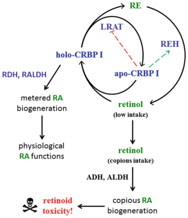
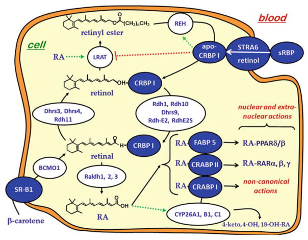
Multiple binding and transport proteins facilitate many aspects of retinoid biology through effects on retinoid transport, cellular uptake, metabolism, and nuclear delivery. These include the serum retinol binding protein sRBP (aka Rbp4), the plasma membrane sRBP receptor Stra6, and the intracellular retinoid binding-proteins such as cellular retinol-binding proteins (CRBP) and cellular retinoic acid binding-proteins (CRABP). sRBP transports the highly lipophilic retinol through an aqueous medium. The major intracellular retinol-binding protein, CRBP1, likely enhances efficient retinoid use by providing a sink to facilitate retinol uptake from sRBP through the plasma membrane or via Stra6, delivering retinol or retinal to select enzymes that generate retinyl esters or retinoic acid, and protecting retinol/retinal from excess catabolism or opportunistic metabolism. Intracellular retinoic acid binding-proteins (CRABP1 and 2, and FABP5) seem to have more diverse functions distinctive to each, such as directing retinoic acid to catabolism, delivering retinoic acid to specific nuclear receptors, and generating non-canonical actions. Gene ablation of intracellular retinoid binding-proteins does not cause embryonic lethality or gross morphological defects. Metabolic and functional defects manifested in knockouts of CRBP1, CRBP2 and CRBP3, however, illustrate their essentiality to health, and in the case of CRBP2, to survival during limited dietary vitamin A. Future studies should continue to address the specific molecular interactions that occur between retinoid binding-proteins and their targets and their precise physiologic contributions to retinoid homeostasis and function.

Keywords: Acyl-CoA:diacylglycerol acyltransferase; Acyl-CoA:monoacylglycerol acyltransferase; Acyl-CoA:retinol acyltransferase; Cellular retinoic acid binding-protein; Cellular retinol binding-protein; Cytochrome P-450; Lecithin:retinol acyltransferase; Peroxisomal proliferator activated receptor δ/β; Retinal; Retinal dehydrogenase; Retinoic acid; Retinoic acid receptor; Retinol; Retinol dehydrogenase; Retinyl ester hydrolase; Serum retinol binding-protein.


Figures

Fig. 2.1
Fig. 2.1
CRBP1 structure. The “worm” diagram at the left shows the two α-helices (green) and the ten anti-parallel β-strands (tan) arranged in two orthogonal sets of five strands each. Light blue denotes links between the strands or strands and helices. Dark blue indicates retinol. Magenta shows selected exterior residues V27, K31 and L35 in αII. Some interior residues also are shown. Between interior residue R58 and the exterior residues are shown (unmarked) from top to bottom L29, F57 and L36. The space-filling diagram in the middle presents a similar perspective as the “worm” diagram, illustrating seclusion of retinol from the cellular milieu. The depiction of partial residues at the right shows a different perspective from that at the far left to reveal more clearly association or relativity close proximity of L29, I32, L36, F57, R58, and I77 with the β-ionone ring of retinol as they point into the interior of CRBP1, and the outward projections of V27, K31 and L35. Structures were generated with the program Cn3D (http://www.ncbi.nlm.nih.gov/Structure/CN3D/cn3d.shtml) with data downloaded from http://www.ncbi.nlm.nih.gov/Structure/mmdb/mmdbsrv.cgi?uid=24299
Fig. 2.2
Fig. 2.2
Kinetics of RE formation by LRAT catalyzed by rat liver microsomes with holo-CRBP substrates. Each reaction was run with CRBP1 in a 1:1 ratio with retinol [192]. The Km values were ~1 and 0.7 μM for holo-CRBP1 and holo-CRBP2, respectively, with adult rat liver microsomes. Use of holo-CRBP did not support RE formation from ARAT activity
Fig. 2.3
Fig. 2.3
Apo-CRBP1 effects on retinol esterification and RE mobilization. Increasing concentrations of apo-CRBP1 increase the hydrolysis of resident RE in liver microsomes, whereas titrating apo-CRBP1 into an LRAT reaction using holo-CRBP1 as substrate inhibits RE formation. These data indicate that the ratio holo-CRBP1/apo-CRBP1 influence flux between retinol and RE. Fully-charged CRBP1 would favor RE formation, while still allowing retinal formation for RA biosynthesis; whereas appreciable apo-CRBP1 would stimulate RE hydrolysis to maintain holo-CRBP1 as substrate for RA biosynthesis
Fig. 2.4
Fig. 2.4
Channeling of intestinal retinal metabolism to direct pro-retinoid and retinoid flux into RE. β-Carotene and pro-vitamin A carotenoids provide the major sources of dietary vitamin A. A dioxygenase (BCO1) generates retinal from β-carotene. Retinal can then be reduced by retinal reductase or dehydrogenated into RA. The dehydrogenation rate of retinal bound to CRBP2 is <300-fold the rate of reduction into retinol. β-Carotene is not toxic, whereas RA is toxic. Moreover, large scale conversion of retinal in the intestine into RA likely would not serve the vitamin A needs of the animal. CRBP2-directed metabolism of retinal through retinol into RE for incorporation into chylomicrons provides a mechanism for efficient conservation of vitamin A as its ester, systemic delivery of RE via chylomicrons, and limiting RA biosynthesis
Fig. 2.5
Fig. 2.5
Disposition of low amounts of dietary retinol vs. copious or toxic dietary retinol. This figure presents a model for retinol toxicity consistent with saturation of retinoid binding-proteins by copious dietary retinol resulting in unmetered generation of RA by xenobiotic clearing enzymes. This path is in contrast to channeling of dietary sufficient amounts of vitamin A by retinoid binding-proteins, such as CRBP1. This path complements the path imposed upon the intestine by CRBP2, which directs dietary retinol into RE, rather than RA
Fig. 2.6
Fig. 2.6
Kinetic relationships between RDH and CRBP1-retinol. The top panel shows the Michaelis-Menten relationship (substrate-concentration dependent, saturable kinetics, initial velocity conditions) between holo-CRBP1 and the rate of retinal formation catalyzed by microsomes. The kinetic constants generated were independent of the ratio total CRBP1/retinol, indicating ability of CRBP1 to deliver retinol without diffusion. The bottom panel shows the impact of mutating external residues of CRBP1 on kinetics of retinal formation using microsomes as source of RDH. The sensitivity of the kinetics to changes in one external CRBP amino acid residue, without affecting affinity for retinol, corroborates an interaction between RDH and CRBP1
Fig. 2.7
Fig. 2.7
Holo-CRBP1 crosslinks with RDH. A. CRBP1 was covalently modified with a heterobi-functional, cleavable crosslinking reagent. The crosslinker was radioiodinated. B. UV irradiation activated the azide of the crosslinker to a nitrene residue that formed a covalent bond between CRBP1 and closely associating RDH (and LRAT). C) Cleavage of the crosslinking reagent left the radioiodine on the target protein, effectively resulting in a transfer of radioiodine from holo-CRBP1 to RDH and LRAT. CRBP1 cross-linked only with two proteins in microsomes, RDH and LRAT, indicating specificity of the interaction. Crosslinking with RDH required presence of NAD(P)+, consistent with the ordered bisubstrate reaction mechanism of a dehydrogenase
Fig. 2.8
Fig. 2.8
Kinetic relationships between CRBP1 and recombinant RALDH1 or RALDH2. Top: conversion of retinal into RA by RALDH1 using CRBP1-retinal (2:1 ratio) or unbound retinal. Middle: Dixon plot of the effect of titrating apo-CRBP1 into 2 μM retinal on generation of RA by RALDH1 illustrating inhibition by apo-CRBP1. These data allow calculating the uninhibited Vm of RALDH1 with CRBP1-retinal, which was ~90 % of the Vm with unbound retinal. The reaction with RAlDH1 was run at 25 °C to reduce its rate to preserve initial velocity conditions. The IC50 of apo-CRBP1 for inhibiting RALDH1 was 1.4 μM. Bottom: conversion of retinal into RA by RALDH2. A twofold excess of binding protein to retinal did not affect the reaction rate, indicating that RALDH2 interacts with CRBP1 [199]
Fig. 2.9
Fig. 2.9
Kinetic relationship between CRABP1 and RA metabolism. Testis microsomes were used as source of RA catabolic activity, because testis has one of the highest intracellular RA concentrations [181]. Kinetics with CRABP1 were generated with a threefold molar excess of CRABP1 to RA to reduce free RA to negligible amounts [68]. Note that the reaction with CRABP1-RA is more efficient than with unbound RA (higher Vm/Km). Note also that the elimination half-life of RA does not change between unbound and CRABP1-bound catabolism, again suggesting interaction between enzyme and binding protein. The rates of RA oxidized at C4 do change, however, suggesting that these bound retinoids would inhibit RA catabolism if in sufficiently high concentrations. With physiological amounts of RA, the 4-oxidized derivatives have very low concentrations, but with toxic amounts of RA, the concentrations of 4-oxidized derivatives increase and contribute to RA toxicity
Fig. 2.10
Fig. 2.10
Model of retinoid homeostasis illustrating roles of retinoid binding-proteins. This model illustrates contributions of retinoid binding-proteins to multiple aspects of retinol uptake and metabolism, and RA catabolism and action. The complexity of storing and activating retinol undoubtedly contributes to the multiplicity of retinoid actions. This chaperoning increases the efficiency of retinoid metabolism, but is not obligatory for life. Note, however, that the CRBP1-null mouse suffers from disrupted retinol metabolism, retinol wasting, and dysfunctions in intermediary metabolism. The dotted lines represent regulatory points

References

    1. Adams MK, Belyaeva OV, Wu L, Kedishvili NY. The retinaldehyde reductase activity of DHRS3 is reciprocally activated by retinol dehydrogenase 10 to control retinoid homeostasis. J Biol Chem. 2014;289:14868–14880. - PMC - PubMed
    1. Ahn T, Kim M, Yun C-H, Chae H-J. Functional regulation of hepatic cytochrome p450 enzymes by physicochemical properties of phospholipids in biological membranes. Curr Protein Pept Sci. 2007;8:496–505. - PubMed
    1. Akerstrom B, Flower DR, Salier JP. Lipocalins: unity in diversity. Biochim Biophys Acta. 2000;1482:1–8. - PubMed
    1. Amengual J, Zhang N, Kemerer M, Maeda T, Palczewski K, Von Lintig J. STRA6 is critical for cellular vitamin A uptake and homeostasis. Hum Mol Genet. 2014 doi: 10.1093/hmg/ddu258. - DOI - PMC - PubMed
    1. Bashor MM, Chytil F. Cellular retinol-binding protein. Biochim Biophys Acta. 1975;411:87–96. - PubMed

MeSH terms