. 2016 Nov 10:6:36447.
doi: 10.1038/srep36447.

ROR1 is a novel prognostic biomarker in patients with lung adenocarcinoma

Affiliations

ROR1 is a novel prognostic biomarker in patients with lung adenocarcinoma

Yu-Zhu Zheng et al. Sci Rep. .

Abstract

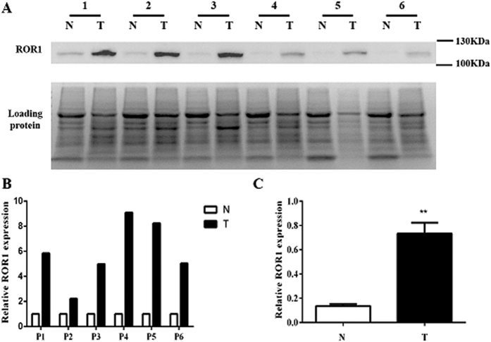
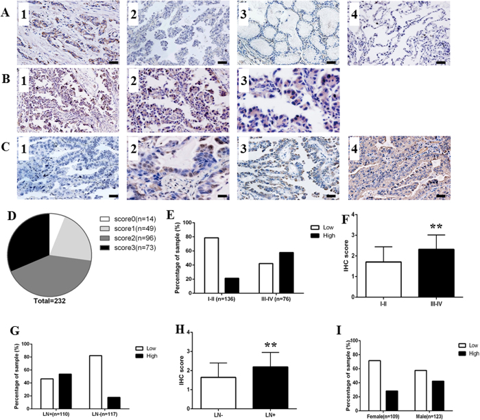
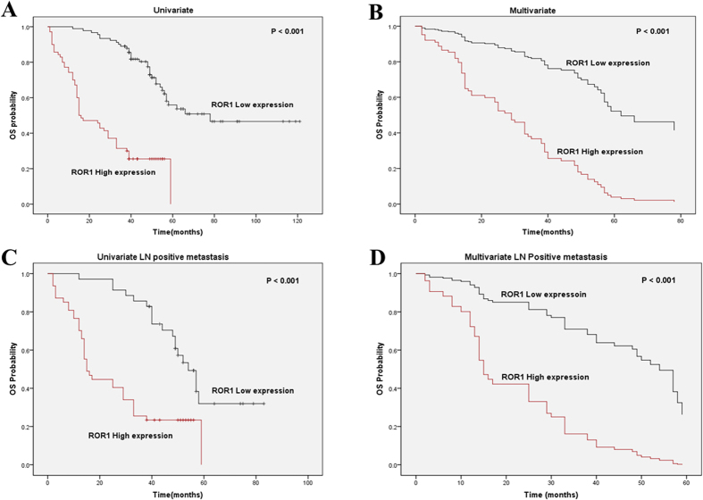
Currently, there is no reliable biomarker to clinically predict the prognosis of lung adenocarcinoma (ADC). The receptor-tyrosine-kinase like orphan receptor 1 (ROR1) is reported to be overexpressed and associated with poor prognosis in several tumors. This study aimed to examine the expression of ROR1 and evaluate its prognostic significance in human lung ADC patients. In this present study, Western blot analysis and immunohistochemistry were performed to characterize expression of ROR1 protein in lung ADC patients. The results revealed that ROR1 protein expression was significantly higher in lung ADC tissues than that in their adjacent non-tumor tissues. Patients at advanced stages and those with positive lymph node metastasis expressed higher level of ROR1 (P < 0.001). Moreover, Chi-square test showed that ROR1 expression was correlated to gender (P = 0.028), the 7th edition of the American Joint Committee on Cancer tumor-node-metastasis (AJCC TNM) staging system and lymph node metastasis (P < 0.001). Kaplan-Meier survival analysis indicated an association of high ROR1 expression with worse overall survival (OS) in lung ADC patients (P < 0.001). Multivariate COX regression analysis further confirmed that ROR1 is an independent prognostic predictor (P < 0.001, HR = 4.114, 95% CI: 2.513-6.375) for OS. Therefore, ROR1 expression significantly correlates with malignant attributes of lung ADC and it may serve as a novel prognostic marker in lung ADC patients.


Figures

Figure 1
Figure 1. Analysis of ROR1 expression in lung ADC and their adjacent non-tumor tissue samples.
(A) Western blot analysis showed that the lung ADC tissue samples (T) express high level of ROR1 protein while the adjacent non-tumor tissue samples (N) express little ROR1 protein. Total protein levels were used as loading control. The full-length gels of Western blot analysis are presented in Supplementary Figure S1. (B,C) The relative intensity of each panel was calculated by Image J and analyzed by paired t-test. Statistics results showed that the value of P < 0.001.
Figure 2
Figure 2. ROR1 expression in lung ADC tissue samples.
(A) The IHC staining of positive and negative control revealed that ROR1 specifically expressed in cancer tissues. Breast cancer tissue stained with anti-ROR1 antibody was used as positive control (1). Negative controls included: lung ADC tissue incubated with PBS (2), nonspecific interstitial pneumonias (3) and adjacent non-tumor tissues (4) stained with anti-ROR1 antibody. (B) The IHC analysis of ROR1 expression in lung ADC tissues showed that ROR1 protein was mainly localized to the cell membrane and cytoplasm of lung ADC cells. Positive staining of ROR1 was shown in brown and the nucleus counterstained with hematoxylin was in blue. The magnification was ×100 in B1, ×200 in B2, ×400 in B3. (C) Different levels of ROR1 expression on TMA detected by IHC analysis. (1) Score 0 indicated that none or little cells express ROR1; (2) score 1 indicated that more than 25% of tumor cells have weak expression of ROR1; (3) score 2 indicated more than 50% of tumor cells have weak expression or more than 25% of tumor cells have moderate expression of ROR1 protein; (4) score 3 indicated more than 75% of tumor cells have moderate expression or more than 50% of tumor cells have strong expression of ROR1. Bar = 100 μm. (D) The pie chart represented the proportion of negative (score 0), weak (score 1), moderate (score 2) and strong staining (score 3) for ROR1 protein of TMA samples. (E) The proportion of low and high staining of ROR1 in lung ADC tissues of different stages was indicated in each bar (P < 0.001). (F) The scores of ROR1 in different stages were analyzed by two tail t-test. Statistical results showed that the value of P < 0.001. (G) The proportion of low and high staining of ROR1 in lung ADC tissues of different lymph node metastasis status was indicated in each bar (P < 0.001). (H) The scores of ROR1 in patients with different status of lymph node metastasis were analyzed by two tail t-test. Statistical results showed that the value of P < 0.001. (I) The proportion of low and high staining of ROR1 in lung ADC tissues of different gender was indicated in each bar (P = 0.023).
Figure 3
Figure 3. High expression of ROR1 is correlated with shorter OS in lung ADC patients and those with positive lymph node metastasis.
(A) Kaplan-Meier survival analysis of ROR1 expression in 161 lung ADC patients split into two groups. Compared to the high ROR1 group, longer OS time was observed in the low ROR1 group. (B) Multivariate Cox regression survival analysis in 161 ADC patients split into two groups. ROR1 expression was determined to be an independent prognostic factor. (C) Kaplan-Meir survival analysis of ROR1 in 82 ADC patients with positive lymph node metastasis. Compared to the high ROR1 expression group, the low ROR1 expression group had better OS time. (D) Multivariate Cox regression survival analysis in 82 ADC patients with positive lymph node metastasis. ROR1 expression was an independent prognostic factor in these patients.

References

    1. Siegel R. L., Miller K. D. & Jemal A. Cancer statistics. CA. Cancer. J. Clin. 66, 7–30 (2016). - PubMed
    1. Lewis D. R., Check D. P., Caporaso N. E., Travis W. D. & Devesa S. S. US lung cancer trends by histologic type. Cancer 120, 2883–2892 (2014). - PMC - PubMed
    1. Li J. et al.. Identification of high-quality cancer prognostic markers and metastasis network modules. Nat Commun. 1, 1–8 (2010). - PMC - PubMed
    1. Sung H. J. & Cho J. Y. Biomarkers for the lung cancer diagnosis and their advances in proteomics. BMB. Rep. 41, 615–625 (2008). - PubMed
    1. van Rens M. T., de la Riviere A. B., Elbers H. R. & van Den Bosch J. M. Prognostic assessment of 2,361 patients who underwent pulmonary resection for non-small cell lung cancer, stage I, II and IIIA. Chest. 117, 374–379 (2000). - PubMed

Publication types

MeSH terms

Substances