

Reed-Sternberg cells in Hodgkin's lymphoma present features of cellular senescence
- PMID: 27831553
- PMCID: PMC5287295
- DOI: 10.1038/cddis.2016.185
Reed-Sternberg cells in Hodgkin's lymphoma present features of cellular senescence
Abstract
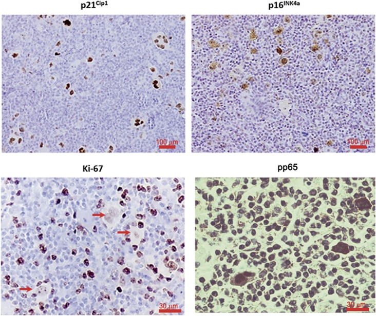
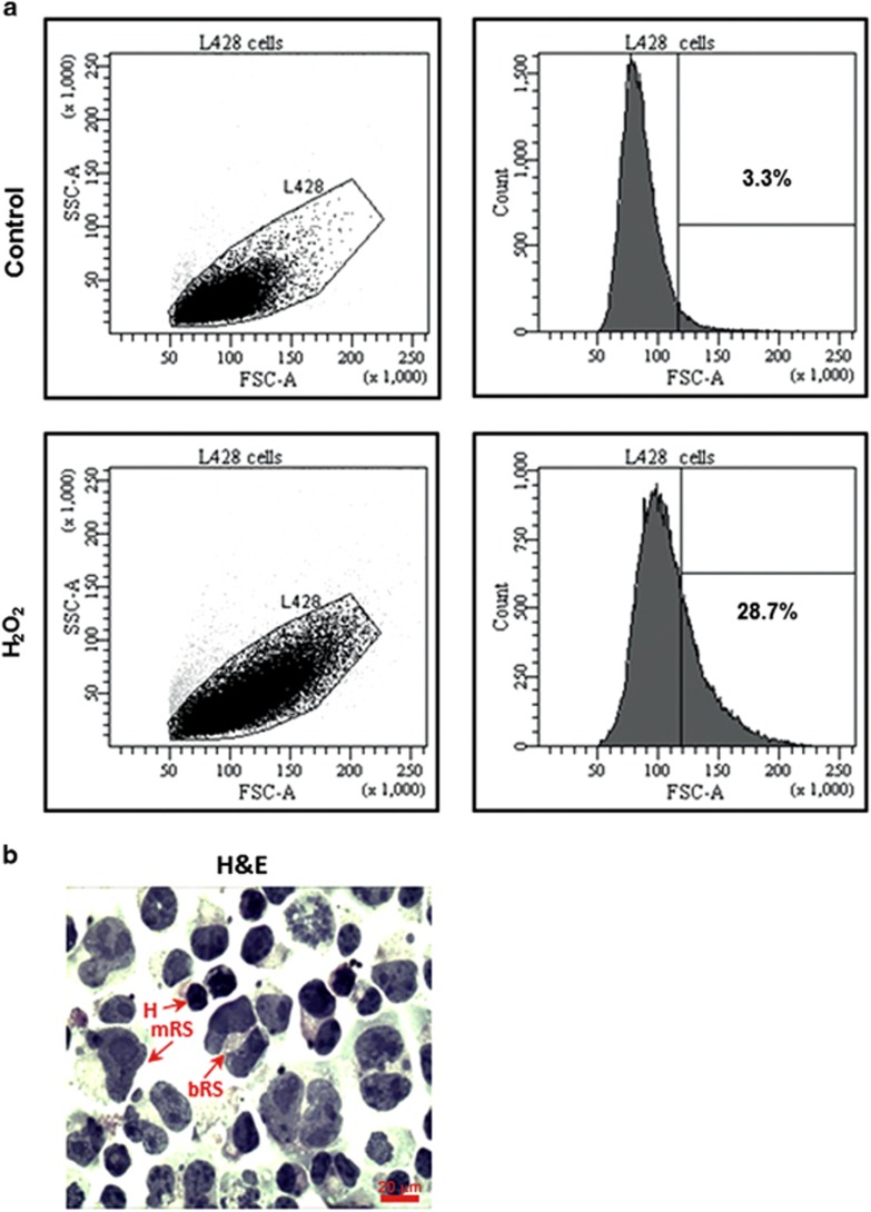
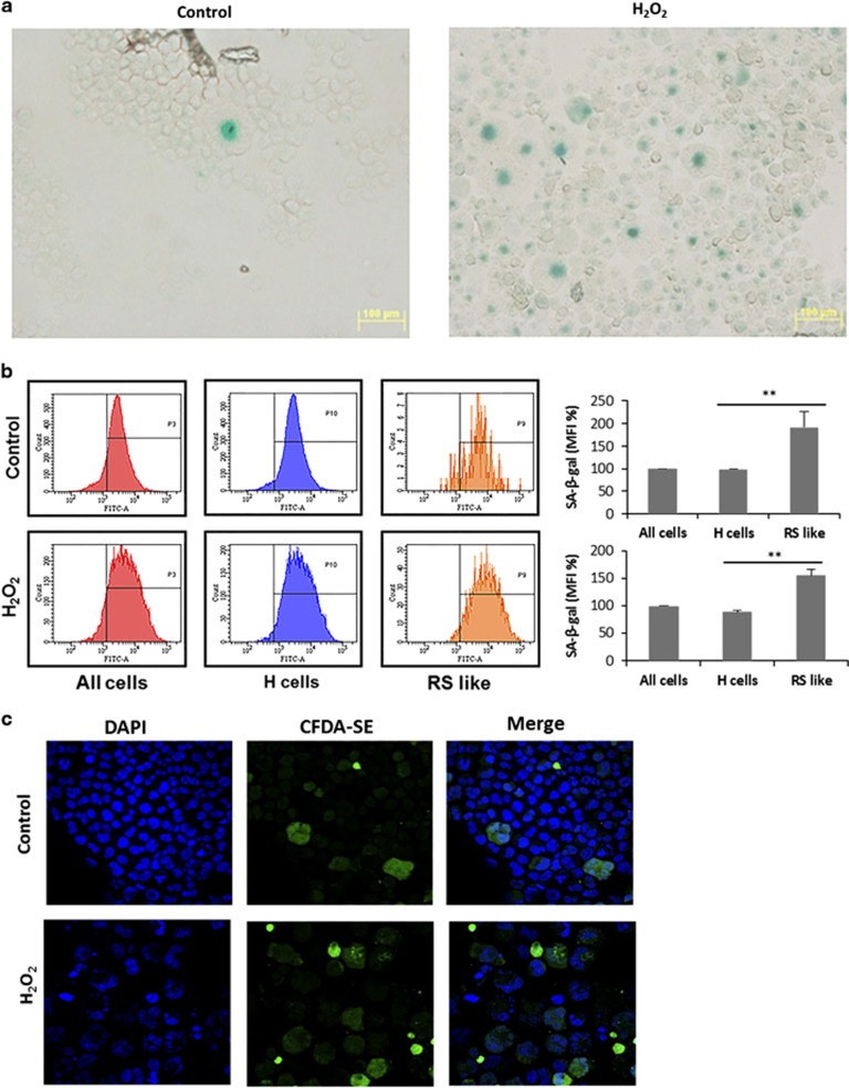
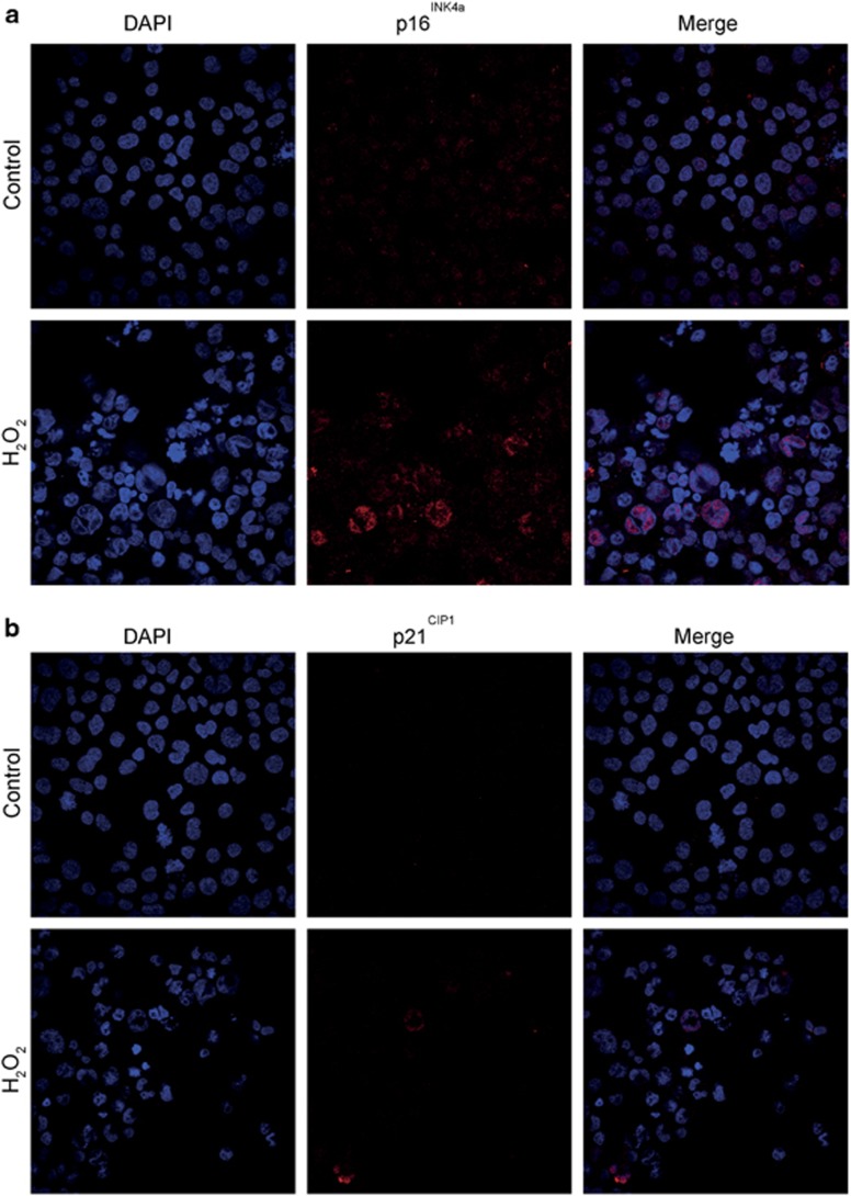
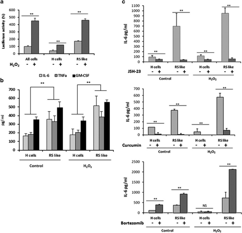
Hodgkin's Lymphoma (HL) is one of the most prevailing malignancies in young adults. Reed-Sternberg (RS) cells in HL have distinctive large cell morphology, are characteristic of the disease and their presence is essential for diagnosis. Enlarged cells are one of the hallmarks of senescence, but whether RS cells are senescent has not been previously investigated. Here we show that RS cells have characteristics of senescent cells; RS cells in HL biopsies specifically express the senescence markers and cell cycle inhibitors p21Cip1 and p16INK4a and are negative for the proliferation marker Ki-67, suggesting that these cells have ceased to proliferate. Moreover, the RS-like cells in HL lines, stained specifically for senescence-associated β-galactosidase (SA-β-gal). Oxidative stress promoted senescence in these cells as demonstrated by their staining for p21Cip1, p16INK4a, p53 and γH2AX. Senescent cells produce copious amounts of inflammatory cytokines termed 'senescence-associated secretory phenotype' (SASP), primarily regulated by Nuclear Factor κB (NF-κB). Indeed, we show that NF-κB activity and NF-κB-dependent cytokines production (e.g., IL-6, TNF-α, GM-CSF) were elevated in RS-like cells. Furthermore, NF-κB inhibitors, JSH-23 and curcumin reduced IL-6 secretion from RS-like cells. Thus, defining RS cells as senescent offers new insights on the origin of the proinflammatory microenvironment in HL.
Figures
Similar articles
-
The side population, as a precursor of Hodgkin and Reed-Sternberg cells and a target for nuclear factor-κB inhibitors in Hodgkin's lymphoma.Cancer Sci. 2010 Nov;101(11):2490-6. doi: 10.1111/j.1349-7006.2010.01693.x. Cancer Sci. 2010. PMID: 20735433 Free PMC article.
-
Interleukin-6, but not interleukin-4, is expressed by Reed-Sternberg cells in Hodgkin's disease with or without histologic features of Castleman's disease.Am J Pathol. 1992 Jul;141(1):129-38. Am J Pathol. 1992. PMID: 1632458 Free PMC article.
-
The molecular basis for the generation of Hodgkin and Reed-Sternberg cells in Hodgkin's lymphoma.Int J Hematol. 2003 May;77(4):330-5. doi: 10.1007/BF02982639. Int J Hematol. 2003. PMID: 12774919 Review.
-
Cytoplasmic aggregation of TRAF2 and TRAF5 proteins in the Hodgkin-Reed-Sternberg cells.Am J Pathol. 2002 May;160(5):1647-54. doi: 10.1016/S0002-9440(10)61112-1. Am J Pathol. 2002. PMID: 12000717 Free PMC article.
-
The biological basis of Hodgkin's lymphoma.Drug News Perspect. 2003 Dec;16(10):649-56. doi: 10.1358/dnp.2003.16.10.829295. Drug News Perspect. 2003. PMID: 14747844 Review.
Cited by
-
Accelerated senescence of bone marrow erythrocyte precursors in myelodysplastic syndrome.Ann Med. 2025 Dec;57(1):2494676. doi: 10.1080/07853890.2025.2494676. Epub 2025 Apr 25. Ann Med. 2025. PMID: 40277030 Free PMC article.
-
Therapy-induced senescent tumor cells in cancer relapse.J Natl Cancer Cent. 2023 Sep 19;3(4):273-278. doi: 10.1016/j.jncc.2023.09.001. eCollection 2023 Dec. J Natl Cancer Cent. 2023. PMID: 39036667 Free PMC article. Review.
-
Mechanisms and significance of therapy-induced and spontaneous senescence of cancer cells.Cell Mol Life Sci. 2020 Jan;77(2):213-229. doi: 10.1007/s00018-019-03261-8. Epub 2019 Aug 14. Cell Mol Life Sci. 2020. PMID: 31414165 Free PMC article. Review.
-
Differentiation of Hodgkin lymphoma cells by reactive oxygen species and regulation by heme oxygenase-1 through HIF-1α.Cancer Sci. 2021 Jun;112(6):2542-2555. doi: 10.1111/cas.14890. Epub 2021 Apr 7. Cancer Sci. 2021. PMID: 33738869 Free PMC article.
-
Rapid Airway Narrowing Associated with Hodgkin's Lymphoma, a Case Report.J Educ Teach Emerg Med. 2020 Apr 15;5(2):V11-V13. doi: 10.21980/J86D3Q. eCollection 2020 Apr. J Educ Teach Emerg Med. 2020. PMID: 37465399 Free PMC article.
References
-
- Boll B, Goergen H, Arndt N, Meissner J, Krause SW, Schnell R et al. Relapsed hodgkin lymphoma in older patients: a comprehensive analysis from the german hodgkin study group. J Clin Oncol 2013; 31: 4431–4437. - PubMed
-
- Kuppers R. New insights in the biology of Hodgkin lymphoma. Hematology Am Soc Hematol Educ Program 2012; 2012: 328–334. - PubMed
-
- Tiacci E, Doring C, Brune V, van Noesel CJ, Klapper W, Mechtersheimer G et al. Analyzing primary Hodgkin and Reed-Sternberg cells to capture the molecular and cellular pathogenesis of classical Hodgkin lymphoma. Blood 2012; 120: 4609–4620. - PubMed
Publication types
MeSH terms
Substances
Grants and funding
LinkOut - more resources
Full Text Sources
Other Literature Sources
Medical
Research Materials
Miscellaneous
