

Manipulating the air-filled zebrafish swim bladder as a neutrophilic inflammation model for acute lung injury
- PMID: 27831560
- PMCID: PMC5260887
- DOI: 10.1038/cddis.2016.365
Manipulating the air-filled zebrafish swim bladder as a neutrophilic inflammation model for acute lung injury
Abstract
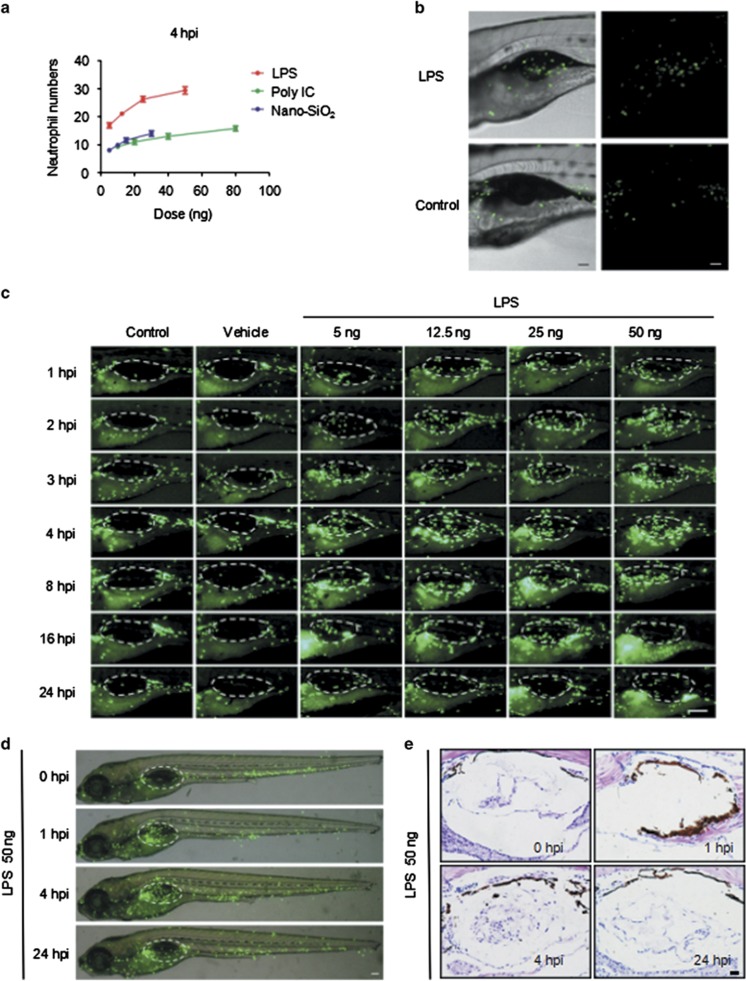
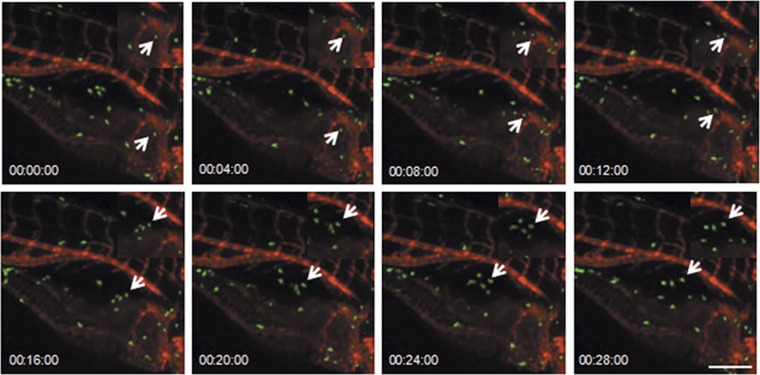
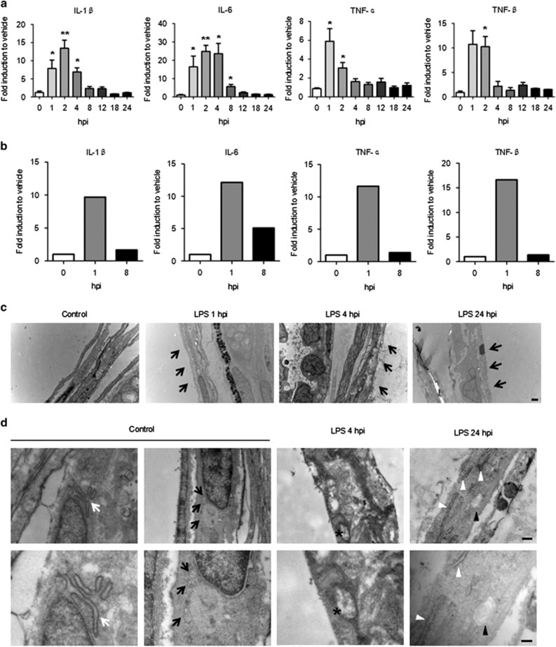
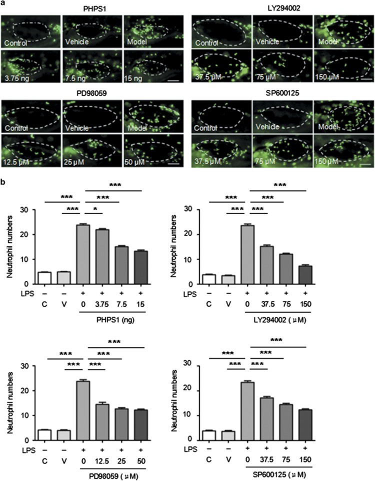
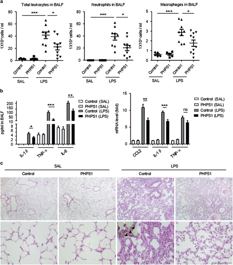
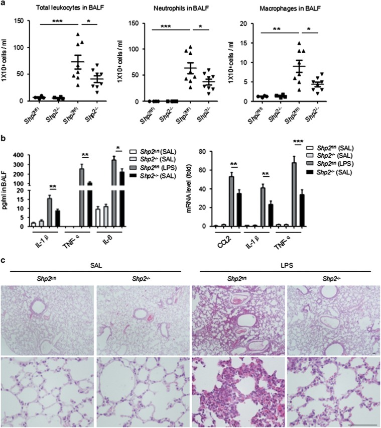
Acute lung injury (ALI) and its more severe form, acute respiratory distress syndrome (ARDS), are life-threatening diseases that are associated with high mortality rates due to treatment limitations. Neutrophils play key roles in the pathogenesis of ALI/ARDS by promoting the inflammation and injury of the alveolar microenvironment. To date, in vivo functional approaches have been limited by the inaccessibility to the alveolar sacs, which are located at the anatomical terminal of the respiratory duct in mammals. We are the first to characterize the swim bladder of the zebrafish larva, which is similar to the mammalian lung, as a real-time in vivo model for examining pulmonary neutrophil infiltration during ALI. We observed that the delivery of exogenous materials, including lipopolysaccharide (LPS), Poly IC and silica nanoparticles, by microinjection triggered significant time- and dose-dependent neutrophil recruitment into the swim bladder. Neutrophils infiltrated the LPS-injected swim bladder through the blood capillaries around the pneumatic duct or a site near the pronephric duct. An increase in the post-LPS inflammatory cytokine mRNA levels coincided with the in vivo neutrophil aggregation in the swim bladder. Microscopic examinations of the LPS-injected swim bladders further revealed in situ injuries, including epithelial distortion, endoplasmic reticulum swelling and mitochondrial injuries. Inhibitor screening assays with this model showed a reduction in neutrophil migration into the LPS-injected swim bladder in response to Shp2 inhibition. Moreover, the pharmacological suppression and targeted disruption of Shp2 in myeloid cells alleviated pulmonary inflammation in the LPS-induced ALI mouse model. Additionally, we used this model to assess pneumonia-induced neutrophil recruitment by microinjecting bronchoalveolar lavage fluid from patients into swim bladders; this injection enhanced neutrophil aggregation relative to the control. In conclusion, our findings highlight the swim bladder as a promising and powerful model for mechanistic and drug screening studies of alveolar injuries.
Figures
Similar articles
-
A novel zebrafish model to emulate lung injury by folate deficiency-induced swim bladder defectiveness and protease/antiprotease expression imbalance.Sci Rep. 2019 Sep 2;9(1):12633. doi: 10.1038/s41598-019-49152-7. Sci Rep. 2019. PMID: 31477754 Free PMC article.
-
Pseudoginsenoside-F11 Attenuates Lipopolysaccharide-Induced Acute Lung Injury by Suppressing Neutrophil Infiltration and Accelerating Neutrophil Clearance.Inflammation. 2019 Oct;42(5):1857-1868. doi: 10.1007/s10753-019-01047-5. Inflammation. 2019. PMID: 31332661
-
Mesenchymal stem cells improves survival in LPS-induced acute lung injury acting through inhibition of NETs formation.J Cell Physiol. 2017 Dec;232(12):3552-3564. doi: 10.1002/jcp.25816. Epub 2017 Feb 9. J Cell Physiol. 2017. PMID: 28112391
-
Deciphering the role of damage-associated molecular patterns and inflammatory responses in acute lung injury.Life Sci. 2022 Sep 15;305:120782. doi: 10.1016/j.lfs.2022.120782. Epub 2022 Jul 6. Life Sci. 2022. PMID: 35809663 Review.
-
Neutrophils in acute lung injury.Front Biosci (Landmark Ed). 2012 Jun 1;17(6):2278-83. doi: 10.2741/4051. Front Biosci (Landmark Ed). 2012. PMID: 22652778 Review.
Cited by
-
Mitochondrial Division Inhibitor 1 Attenuates Mitophagy in a Rat Model of Acute Lung Injury.Biomed Res Int. 2019 May 8;2019:2193706. doi: 10.1155/2019/2193706. eCollection 2019. Biomed Res Int. 2019. PMID: 31205936 Free PMC article.
-
Application of transgenic zebrafish for investigating inflammatory responses to nanomaterials: Recommendations for new users.F1000Res. 2025 Jun 23;12:51. doi: 10.12688/f1000research.128851.2. eCollection 2023. F1000Res. 2025. PMID: 40656220 Free PMC article. Review.
-
Targeting protein phosphatases for the treatment of inflammation-related diseases: From signaling to therapy.Signal Transduct Target Ther. 2022 Jun 4;7(1):177. doi: 10.1038/s41392-022-01038-3. Signal Transduct Target Ther. 2022. PMID: 35665742 Free PMC article. Review.
-
A review on current advancement in zebrafish models to study chronic inflammatory diseases and their therapeutic targets.Heliyon. 2024 May 23;10(11):e31862. doi: 10.1016/j.heliyon.2024.e31862. eCollection 2024 Jun 15. Heliyon. 2024. PMID: 38867970 Free PMC article. Review.
-
Inflammatory, Oxidative Stress, and Apoptosis Effects in Zebrafish Larvae after Rapid Exposure to a Commercial Glyphosate Formulation.Biomedicines. 2021 Nov 27;9(12):1784. doi: 10.3390/biomedicines9121784. Biomedicines. 2021. PMID: 34944599 Free PMC article.
References
-
- Ware LB, Matthay MA. The acute respiratory distress syndrome. N Engl J Med 2000; 342: 1334–1349. - PubMed
-
- Gong MN, Thompson BT. Acute respiratory distress syndrome: shifting the emphasis from treatment to prevention. Curr Opin Crit Care 2016; 22: 21–37. - PubMed
-
- Ranieri VM, Rubenfeld GD, Thompson BT, Ferguson ND, Caldwell E, Fan E et al. Acute respiratory distress syndrome: the Berlin definition. JAMA 2012; 307: 2526–2533. - PubMed
-
- Zaas AK, Schwartz DA. Innate immunity and the lung: defense at the interface between host and environment. Trends Cardiovas Med 2005; 15: 195–202. - PubMed
Publication types
MeSH terms
Substances
LinkOut - more resources
Full Text Sources
Other Literature Sources
Molecular Biology Databases
