

A Biomechanical Modeling Guided CBCT Estimation Technique
- PMID: 27831866
- PMCID: PMC5381525
- DOI: 10.1109/TMI.2016.2623745
A Biomechanical Modeling Guided CBCT Estimation Technique
Abstract
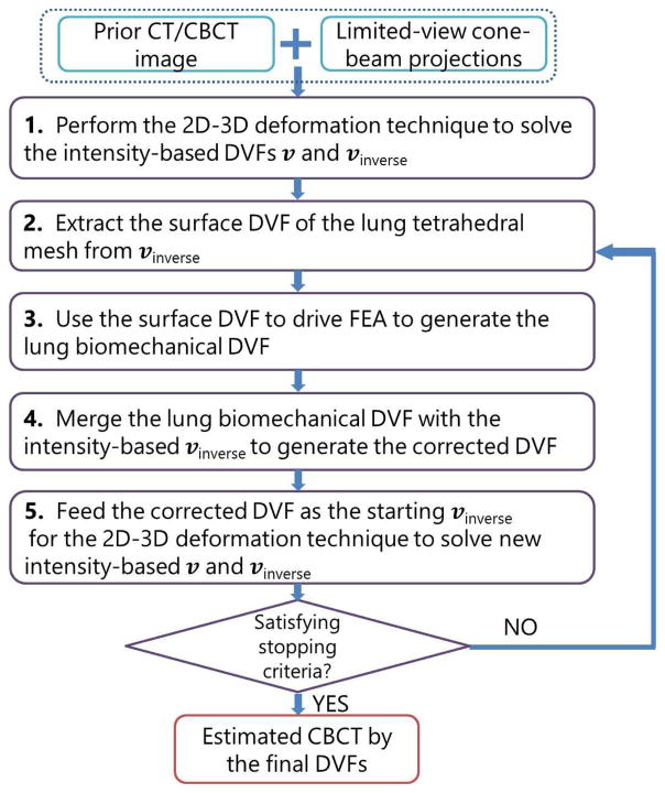
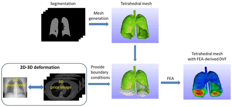
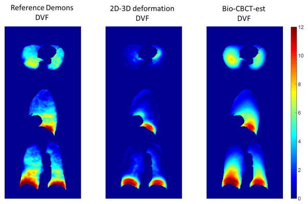
Two-dimensional-to-three-dimensional (2D-3D) deformation has emerged as a new technique to estimate cone-beam computed tomography (CBCT) images. The technique is based on deforming a prior high-quality 3D CT/CBCT image to form a new CBCT image, guided by limited-view 2D projections. The accuracy of this intensity-based technique, however, is often limited in low-contrast image regions with subtle intensity differences. The solved deformation vector fields (DVFs) can also be biomechanically unrealistic. To address these problems, we have developed a biomechanical modeling guided CBCT estimation technique (Bio-CBCT-est) by combining 2D-3D deformation with finite element analysis (FEA)-based biomechanical modeling of anatomical structures. Specifically, Bio-CBCT-est first extracts the 2D-3D deformation-generated displacement vectors at the high-contrast anatomical structure boundaries. The extracted surface deformation fields are subsequently used as the boundary conditions to drive structure-based FEA to correct and fine-tune the overall deformation fields, especially those at low-contrast regions within the structure. The resulting FEA-corrected deformation fields are then fed back into 2D-3D deformation to form an iterative loop, combining the benefits of intensity-based deformation and biomechanical modeling for CBCT estimation. Using eleven lung cancer patient cases, the accuracy of the Bio-CBCT-est technique has been compared to that of the 2D-3D deformation technique and the traditional CBCT reconstruction techniques. The accuracy was evaluated in the image domain, and also in the DVF domain through clinician-tracked lung landmarks.
Figures


Similar articles
-
4D liver tumor localization using cone-beam projections and a biomechanical model.Radiother Oncol. 2019 Apr;133:183-192. doi: 10.1016/j.radonc.2018.10.040. Epub 2018 Nov 14. Radiother Oncol. 2019. PMID: 30448003 Free PMC article.
-
U-net-based deformation vector field estimation for motion-compensated 4D-CBCT reconstruction.Med Phys. 2020 Jul;47(7):3000-3012. doi: 10.1002/mp.14150. Epub 2020 Apr 27. Med Phys. 2020. PMID: 32198934
-
Enhancing liver tumor localization accuracy by prior-knowledge-guided motion modeling and a biomechanical model.Quant Imaging Med Surg. 2019 Jul;9(7):1337-1349. doi: 10.21037/qims.2019.07.04. Quant Imaging Med Surg. 2019. PMID: 31448218 Free PMC article.
-
Automatic liver tumor localization using deep learning-based liver boundary motion estimation and biomechanical modeling (DL-Bio).Med Phys. 2021 Dec;48(12):7790-7805. doi: 10.1002/mp.15275. Epub 2021 Nov 19. Med Phys. 2021. PMID: 34632589 Free PMC article.
-
Three dimensional (3D) imaging techniques in orthodontics-An update.J Family Med Prim Care. 2020 Jun 30;9(6):2626-2630. doi: 10.4103/jfmpc.jfmpc_64_20. eCollection 2020 Jun. J Family Med Prim Care. 2020. PMID: 32984098 Free PMC article. Review.
Cited by
-
Deep Filtered Back Projection for CT Reconstruction.IEEE Access. 2024;12:20962-20972. doi: 10.1109/access.2024.3357355. Epub 2024 Jan 22. IEEE Access. 2024. PMID: 39211346 Free PMC article.
-
A biomechanical modeling-guided simultaneous motion estimation and image reconstruction technique (SMEIR-Bio) for 4D-CBCT reconstruction.Phys Med Biol. 2018 Feb 8;63(4):045002. doi: 10.1088/1361-6560/aaa730. Phys Med Biol. 2018. PMID: 29328048 Free PMC article.
-
Statistical Iterative CBCT Reconstruction Based on Neural Network.IEEE Trans Med Imaging. 2018 Jun;37(6):1511-1521. doi: 10.1109/TMI.2018.2829896. IEEE Trans Med Imaging. 2018. PMID: 29870378 Free PMC article.
-
Clinical Study of Orthogonal-View Phase-Matched Digital Tomosynthesis for Lung Tumor Localization.Technol Cancer Res Treat. 2017 Dec;16(6):866-878. doi: 10.1177/1533034617705716. Epub 2017 Apr 28. Technol Cancer Res Treat. 2017. PMID: 28449625 Free PMC article.
-
Finite element analysis of a one-piece zirconia implant in anterior single tooth implant applications.PLoS One. 2020 Feb 24;15(2):e0229360. doi: 10.1371/journal.pone.0229360. eCollection 2020. PLoS One. 2020. PMID: 32092128 Free PMC article.
References
-
- Xing L, et al. Overview of image-guided radiation therapy. Med Dosim. 2006 Summer;31:91–112. - PubMed
-
- Kim DW, Chung WK, Yoon M. Imaging doses and secondary cancer risk from kilovoltage cone-beam CT in radiation therapy. Health Phys. 2013 May;104:499–503. - PubMed
-
- Li R, et al. Real-time volumetric image reconstruction and 3D tumor localization based on a single x-ray projection image for lung cancer radiotherapy. Med Phys. 2010 Jun;37:2822–6. - PubMed
-
- Ren L, et al. Development and clinical evaluation of a three-dimensional cone-beam computed tomography estimation method using a deformation field map. Int J Radiat Oncol Biol Phys. 2012 Apr 1;82:1584–93. - PubMed
MeSH terms
Grants and funding
LinkOut - more resources
Full Text Sources
Other Literature Sources
Research Materials
