Review
. 2016 Nov 10;10(11):e0004983.
doi: 10.1371/journal.pntd.0004983. eCollection 2016 Nov.

Comparative Bioinformatics Analysis of Transcription Factor Genes Indicates Conservation of Key Regulatory Domains among Babesia bovis, Babesia microti, and Theileria equi

Affiliations
Review

Comparative Bioinformatics Analysis of Transcription Factor Genes Indicates Conservation of Key Regulatory Domains among Babesia bovis, Babesia microti, and Theileria equi

Heba F Alzan et al. PLoS Negl Trop Dis. .

Abstract

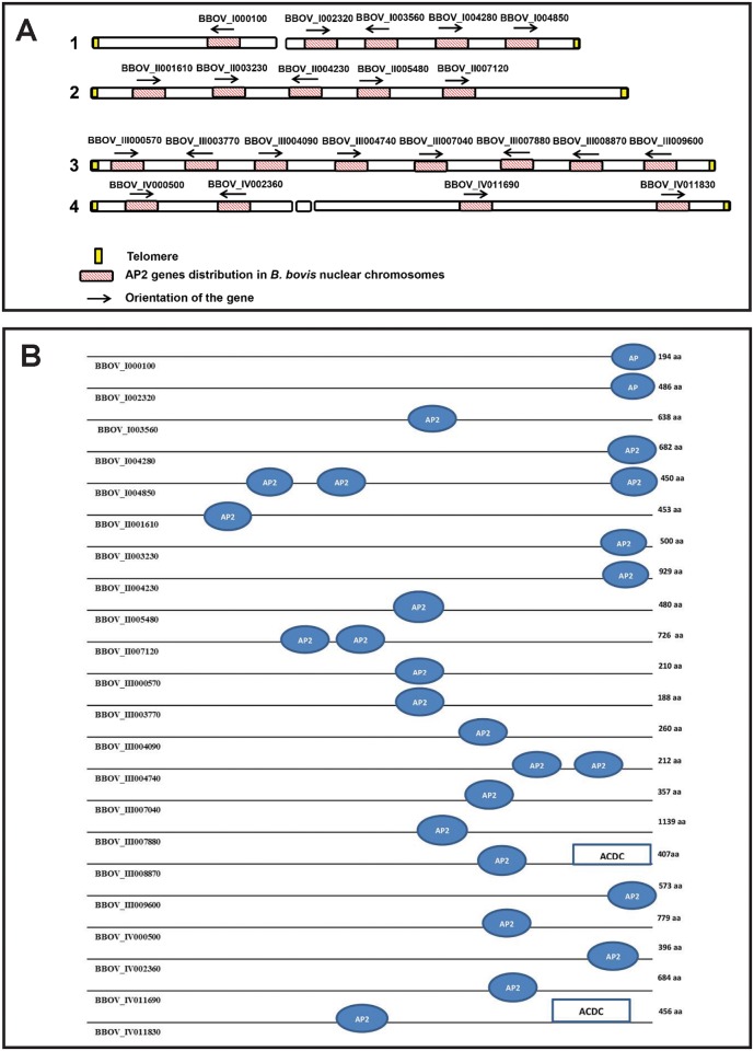
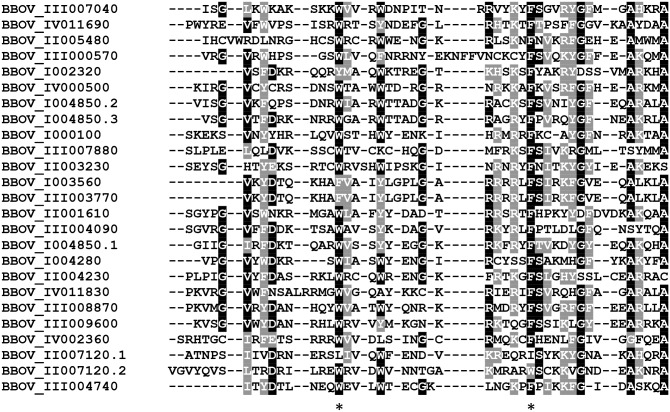
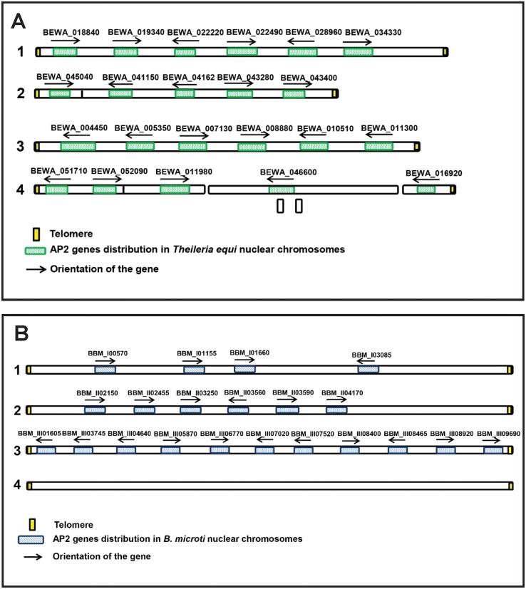
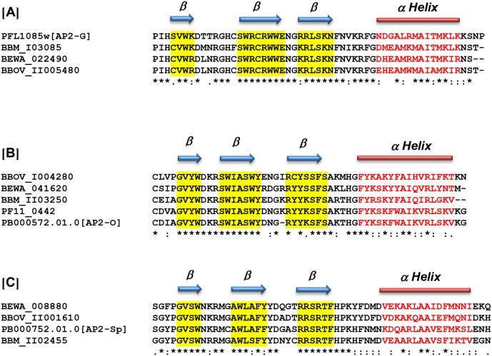
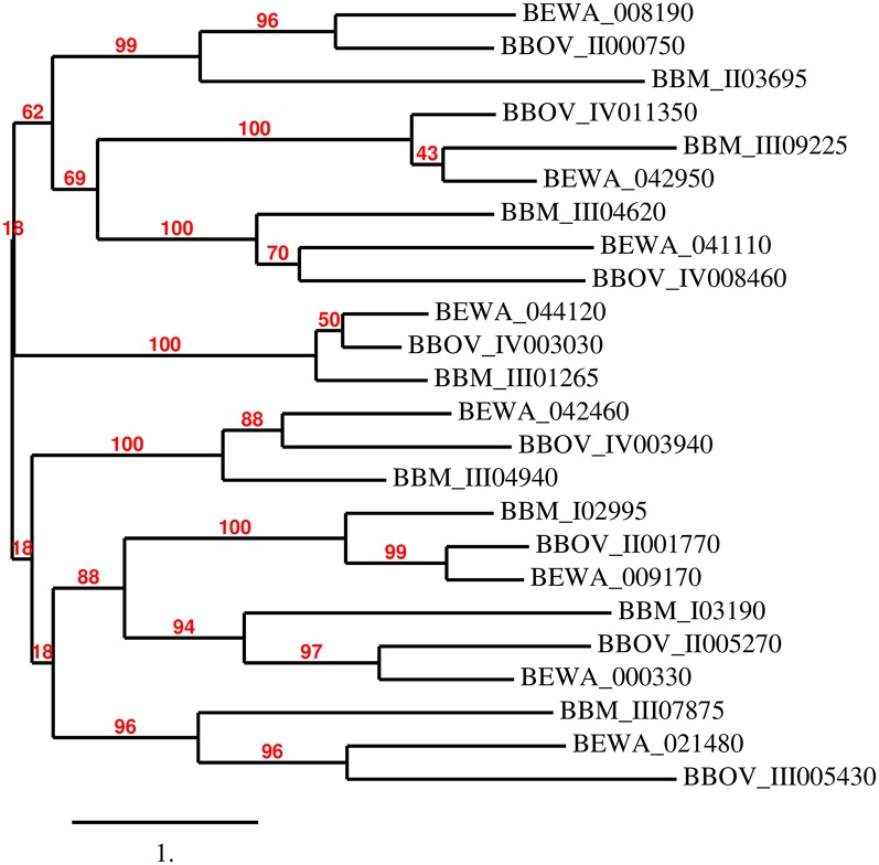
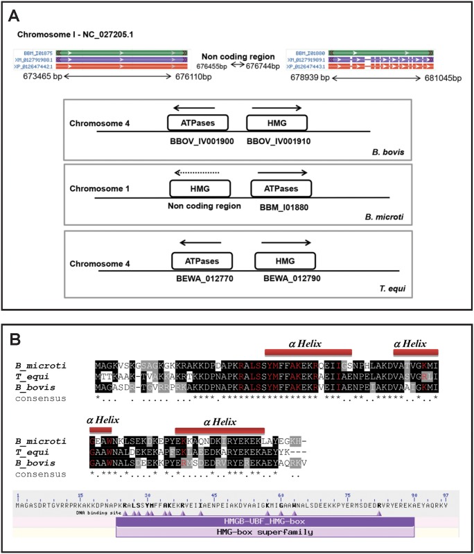
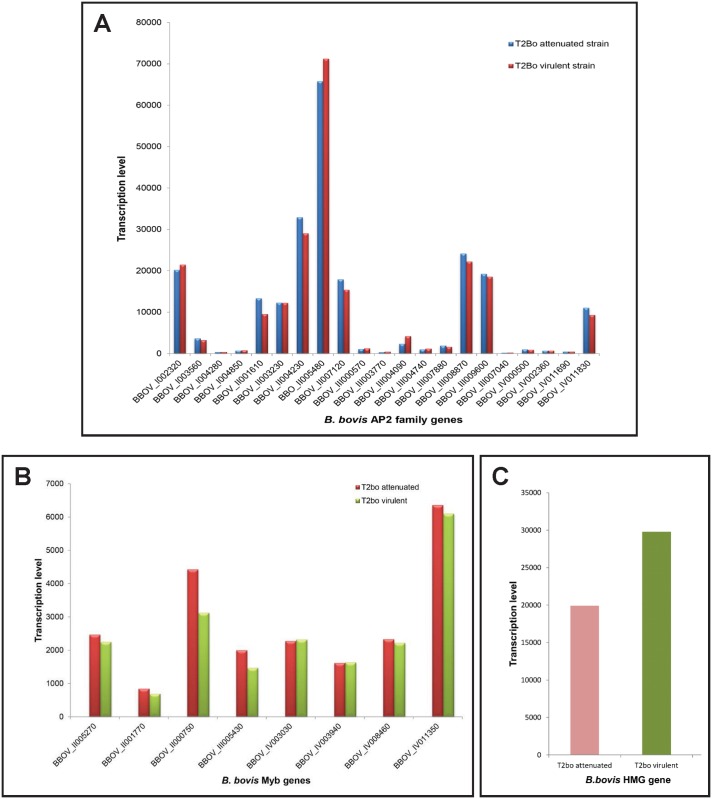
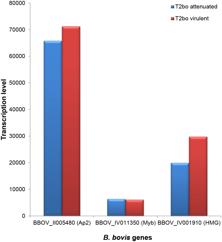
Apicomplexa tick-borne hemoparasites, including Babesia bovis, Babesia microti, and Theileria equi are responsible for bovine and human babesiosis and equine theileriosis, respectively. These parasites of vast medical, epidemiological, and economic impact have complex life cycles in their vertebrate and tick hosts. Large gaps in knowledge concerning the mechanisms used by these parasites for gene regulation remain. Regulatory genes coding for DNA binding proteins such as members of the Api-AP2, HMG, and Myb families are known to play crucial roles as transcription factors. Although the repertoire of Api-AP2 has been defined and a HMG gene was previously identified in the B. bovis genome, these regulatory genes have not been described in detail in B. microti and T. equi. In this study, comparative bioinformatics was used to: (i) identify and map genes encoding for these transcription factors among three parasites' genomes; (ii) identify a previously unreported HMG gene in B. microti; (iii) define a repertoire of eight conserved Myb genes; and (iv) identify AP2 correlates among B. bovis and the better-studied Plasmodium parasites. Searching the available transcriptome of B. bovis defined patterns of transcription of these three gene families in B. bovis erythrocyte stage parasites. Sequence comparisons show conservation of functional domains and general architecture in the AP2, Myb, and HMG proteins, which may be significant for the regulation of common critical parasite life cycle transitions in B. bovis, B. microti, and T. equi. A detailed understanding of the role of gene families encoding DNA binding proteins will provide new tools for unraveling regulatory mechanisms involved in B. bovis, B. microti, and T. equi life cycles and environmental adaptive responses and potentially contributes to the development of novel convergent strategies for improved control of babesiosis and equine piroplasmosis.


Conflict of interest statement

The authors have declared that no competing interests exist.

Figures

Fig 1
Fig 1. Schematic representation of the location of the 22 AP2 genes and the number and domains identified in the genome of B. bovis (data not presented to scale).
(A) The figure represents the genome localization of the B. bovis AP2 genes, distributed among the four chromosomes. Each gene orientation is indicated by black arrows. (B) Gene nomenclature, schematic representation of the number and location of the AP2 domains of the B. bovis AP2 proteins. The presence and location of the ACDC domains in the protein are also indicated.
Fig 2
Fig 2. Sequence alignment of all AP2 domains found in the 22 B. bovis AP2 proteins.
Gene denominations are indicated on the left. Numerations (1–3) indicate the number of domains in each protein. Similar and identical amino acid residues are indicated in gray and black font, respectively. The [*] indicates conservation of W and F amino acids residues among the B. bovis AP2 domains.
Fig 3
Fig 3
(A) Schematic representation of the location of the 22 AP2 genes identified in the genome of T. equi (data not presented in scale). (B) Schematic representation of the location of the 21 AP2 genes identified in the genome of B. microti (data not presented in scale).
Fig 4
Fig 4. Alignments among B. bovis, B. microti, T equi and functionally defined Plasmodium Ap2 domains.
Alignments of: (A) B. bovis BBOV_II005480, B. microti BBM_I03085, and T. equi BEWA_022490 with P. berghei PB000752.01.0, a domain involved in the development of sexual-stage forms in Plasmodium (AP2-G). (B) AP2 domain of the protein encoded by the B. bovis AP2 gene BBOV_I004280, B. microti BBM_II03250, and T. equi BEWA_041620 with putative orthologues in P. berghei (PB00572.01) and P. falciparum, (PF11_0442). Both Plasmodium AP2 proteins are required for the development of ookinetes, and known as AP2-O. (C) B. bovis BBOV_II001610, B. microti BBM_II02455, and T. equi BEWA_008880 with P. berghei PB000752.01.0, a domain involved in the development of sporozoites (AP2-Sp). Predicted secondary structures for the domains of interest are depicted at the top of each of the alignments.
Fig 5
Fig 5. Phylogenetic relationships among the putative Myb proteins of B. bovis, T. equi, and B. microti.
The unrooted phylogenetic tree was generated using the sequences of all the putative Myb proteins identified in B. bovis, B. microti, and T. equi with the program phylogeny.fr. The program calculated the branch support values in percent (%, red font) using an aLRT statistical test (http://www.phylogeny.fr/simple_phylogeny.cgi?workflow_id=ddf5cfc42f5b6c10f3df67f5152bf59a&tab_index=6&go_next=1#anchor).
Fig 6
Fig 6. Putative HMG proteins and genes of B. bovis, T. equi, and B. microti.
(A) Upper: Schematic representation of non-coding area in B. microti genome between 676455 bp and 676744 bp in chromosome 1. Lower: Synteny map for the HMG genes in the B. bovis, B. microti, and T. equi. (B) Upper: Alignment of the deduced amino acid sequences of HMG proteins identified in B. bovis (BBOV_IV001910), T. equi (BEWA_012790), and B. microti (no gene assignment). The residues in red represent the amino acids defining the HMG domain Lower: Location of the HMG domain and depiction of the amino acid residues defining the HMG domain (bold fonts) in the HMG protein encoded by the B. bovis gene BBOV_ IV001910.
Fig 7
Fig 7. Normalized transcriptional profiles of B. bovis AP2 genes using microarray analysis in attenuated and virulent B. bovis strains.
(A) Microarray transcriptional levels expressed as relative transcription units log cpm (copy per million) are represented in the y-axis, and denominations for each B. bovis AP2 gene are represented in the x-axis. (B) Transcription profile of the eight Myb genes identified in the virulent and attenuated T2bo strains of B. bovis using microarray analysis. (C) Transcription profile of the BBOV_IV001910 HMG gene by microarray analysis in virulent and attenuated T2Bo strain of B. bovis. The transcriptome analysis was performed in triplicate on full blood stages of parasites developing in asynchronous cultures.
Fig 8
Fig 8. Comparison among the highest transcribed genes of the AP2, Myb, and HMG genes of B. bovis.
The Ap2 gene BBOV_II005480 shows the highest transcription levels compared to the highest transcribed genes of the Myb and HMG gene groups in B. bovis.

Similar articles

Cited by

References

    1. Scoles GA, Ueti MW. Vector ecology of equine piroplasmosis. Annual review of entomology. 2015;60:561–80. Epub 2015/01/08. 10.1146/annurev-ento-010814-021110 . - DOI - PubMed
    1. Suarez CE, Noh S. Emerging perspectives in the research of bovine babesiosis and anaplasmosis. Vet Parasitol. 2011;180(1–2):109–25. Epub 2011/06/21. 10.1016/j.vetpar.2011.05.032 . - DOI - PubMed
    1. Brayton KA, Lau AO, Herndon DR, Hannick L, Kappmeyer LS, Berens SJ, et al. Genome sequence of Babesia bovis and comparative analysis of apicomplexan hemoprotozoa. PLoS Pathog. 2007;3(10):e148. - PMC - PubMed
    1. Cornillot E, Hadj-Kaddour K, Dassouli A, Noel B, Ranwez V, Vacherie B, et al. Sequencing of the smallest Apicomplexan genome from the human pathogen Babesia microti. Nucleic acids research. 2012;40(18):9102–14. 10.1093/nar/gks700 - DOI - PMC - PubMed
    1. Cornillot E, Dassouli A, Garg A, Pachikara N, Randazzo S, Depoix D, et al. Whole genome mapping and re-organization of the nuclear and mitochondrial genomes of Babesia microti isolates. PloS one. 2013;8(9):e72657 10.1371/journal.pone.0072657 - DOI - PMC - PubMed

MeSH terms