

Microplasma Induced Cell Morphological Changes and Apoptosis of Ex Vivo Cultured Human Anterior Lens Epithelial Cells - Relevance to Capsular Opacification
- PMID: 27832099
- PMCID: PMC5104483
- DOI: 10.1371/journal.pone.0165883
Microplasma Induced Cell Morphological Changes and Apoptosis of Ex Vivo Cultured Human Anterior Lens Epithelial Cells - Relevance to Capsular Opacification
Abstract
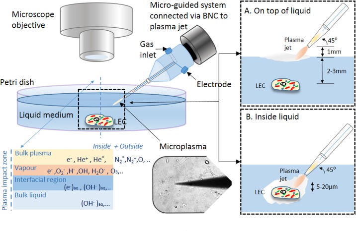
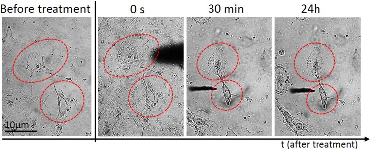
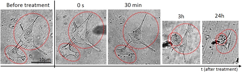
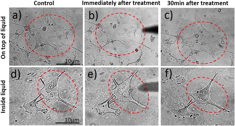
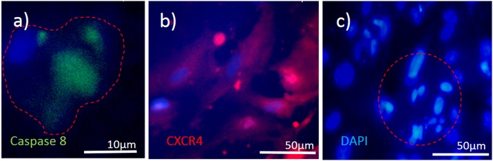
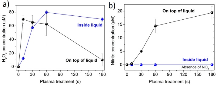
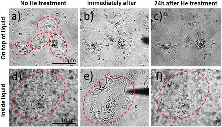
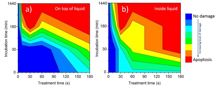
Inducing selective or targeted cell apoptosis without affecting large number of neighbouring cells remains a challenge. A plausible method for treatment of posterior capsular opacification (PCO) due to remaining lens epithelial cells (LECs) by reactive chemistry induced by localized single electrode microplasma discharge at top of a needle-like glass electrode with spot size ~3 μm is hereby presented. The focused and highly-localized atmospheric pressure microplasma jet with electrode discharge could induce a dose-dependent apoptosis in selected and targeted individual LECs, which could be confirmed by real-time monitoring of the morphological and structural changes at cellular level. Direct cell treatment with microplasma inside the medium appeared more effective in inducing apoptosis (caspase 8 positivity and DNA fragmentation) at a highly targeted cell level compared to treatment on top of the medium (indirect treatment). Our results show that single cell specific micropipette plasma can be used to selectively induce demise in LECs which remain in the capsular bag after cataract surgery and thus prevent their migration (CXCR4 positivity) to the posterior lens capsule and PCO formation.
Conflict of interest statement
The authors have declared that no competing interests exist.
Figures
Similar articles
-
Morphological and proliferative studies on ex vivo cultured human anterior lens epithelial cells - relevance to capsular opacification.Acta Ophthalmol. 2015 Sep;93(6):e499-506. doi: 10.1111/aos.12655. Epub 2015 Jan 28. Acta Ophthalmol. 2015. PMID: 25631167
-
Selenium functionalized intraocular lenses inhibit posterior capsule opacification in an ex vivo canine lens capsular bag assay.Exp Eye Res. 2009 Nov;89(5):728-34. doi: 10.1016/j.exer.2009.06.016. Epub 2009 Jul 5. Exp Eye Res. 2009. PMID: 19583956
-
Experimental lens capsular bag model for posterior capsule opacification.Cell Tissue Res. 2014 Jul;357(1):101-8. doi: 10.1007/s00441-014-1870-4. Epub 2014 May 6. Cell Tissue Res. 2014. PMID: 24793776
-
Prevention of posterior capsular opacification.Exp Eye Res. 2015 Jul;136:100-15. doi: 10.1016/j.exer.2015.03.011. Epub 2015 Mar 14. Exp Eye Res. 2015. PMID: 25783492 Review.
-
Experimental models for posterior capsule opacification research.Exp Eye Res. 2016 Jan;142:2-12. doi: 10.1016/j.exer.2015.04.021. Epub 2015 May 1. Exp Eye Res. 2016. PMID: 25939555 Review.
Cited by
-
Direct and Indirect Bactericidal Effects of Cold Atmospheric-Pressure Microplasma and Plasma Jet.Molecules. 2021 Apr 26;26(9):2523. doi: 10.3390/molecules26092523. Molecules. 2021. PMID: 33925959 Free PMC article.
-
Direct Applications of Non-Thermal Atmospheric Pressure Plasma: An Emerging Therapeutic Era in Ophthalmology.Clin Ophthalmol. 2024 May 30;18:1555-1562. doi: 10.2147/OPTH.S462228. eCollection 2024. Clin Ophthalmol. 2024. PMID: 38832076 Free PMC article. Review.
-
Methodologies to unlock the molecular expression and cellular structure of ocular lens epithelial cells.Front Cell Dev Biol. 2022 Sep 13;10:983178. doi: 10.3389/fcell.2022.983178. eCollection 2022. Front Cell Dev Biol. 2022. PMID: 36176273 Free PMC article.
-
Anti-Tumor Activity of Cembranoid-Type Diterpenes Isolated from Nicotiana tabacum L.Biomolecules. 2019 Jan 28;9(2):45. doi: 10.3390/biom9020045. Biomolecules. 2019. PMID: 30696084 Free PMC article.
-
Three-Dimensional Human Cell Culture Models to Study the Pathophysiology of the Anterior Eye.Pharmaceutics. 2020 Dec 15;12(12):1215. doi: 10.3390/pharmaceutics12121215. Pharmaceutics. 2020. PMID: 33333869 Free PMC article. Review.
References
-
- Morfill GE, Kong MG, Zimmermann JL. Focus on plasma medicine. New J Phys. 2009;11 10.1088/1367-2630/11/11/115011 - DOI
-
- Isbary G, Stolz W, Shimizu T, Monetti R, Bunk W, Schmidt HU, et al. Cold atmospheric argon plasma treatment may accelerate wound healing in chronic wounds: Results of an open retrospective randomized controlled study in vivo. Clinical Plasma Medicine. 2013;1(2):25–30. 10.1016/j.cpme.2013.06.001. - DOI
-
- Lazović S, Puač N, Miletić M, Pavlica D, Jovanović M, Bugarski D, et al. The effect of a plasma needle on bacteria in planktonic samples and on peripheral blood mesenchymal stem cells. New J Phys. 2010;12 10.1088/1367-2630/12/8/083037 - DOI
MeSH terms
Substances
LinkOut - more resources
Full Text Sources
Other Literature Sources
