

Vitrectomy and scleral imbrication in patients with myopic traction maculopathy and macular hole retinal detachment
- PMID: 27832339
- PMCID: PMC5364242
- DOI: 10.1007/s00417-016-3523-7
Vitrectomy and scleral imbrication in patients with myopic traction maculopathy and macular hole retinal detachment
Abstract
Purpose: To determine the outcomes of vitrectomy with scleral imbrication in highly myopic eyes with either myopic traction maculopathy (MTM) or macular hole retinal detachment (MHRD).
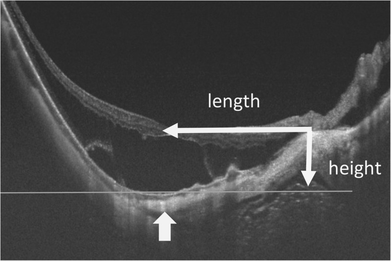
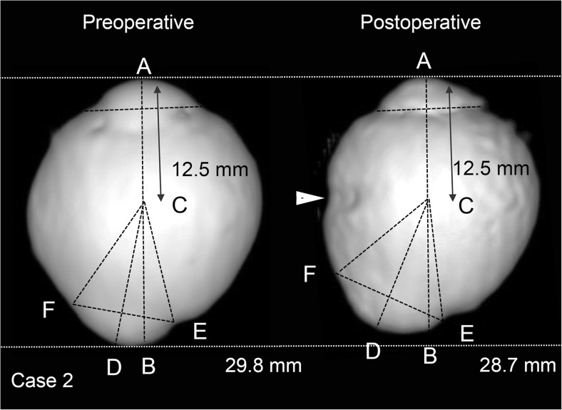
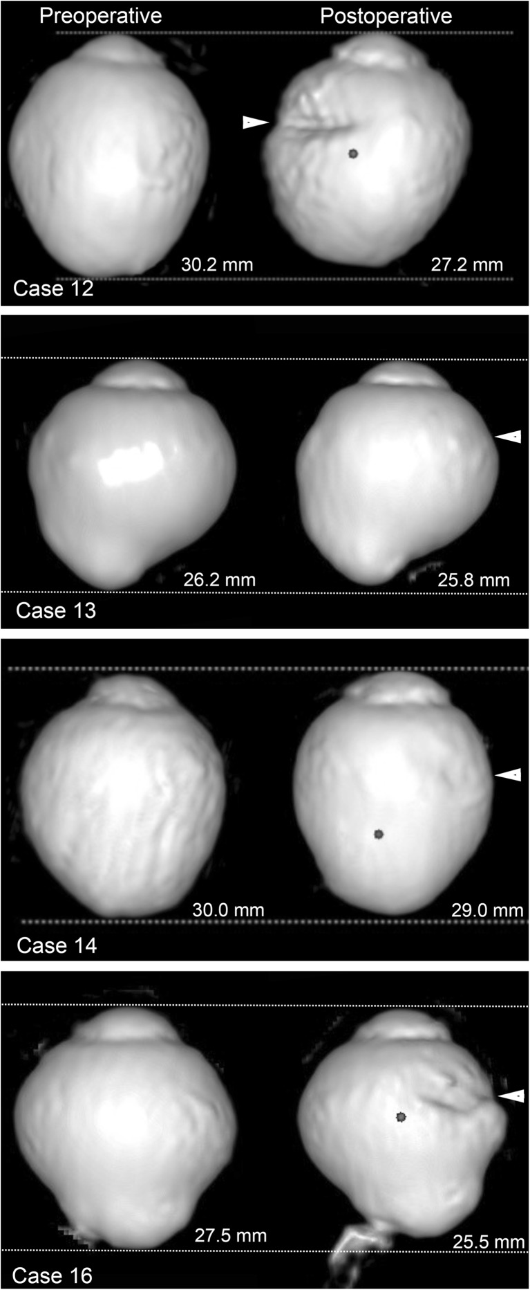
Methods: The medical records of 17 patients who had undergone vitrectomy with internal limiting membrane (ILM) peeling and scleral imbrication for MTM or MHRD were reviewed. The best-corrected visual acuities (BCVAs), the axial length, the macular hole (MH) closure rate, and the shape of the posterior segment determined by optical coherence tomography were evaluated. Three-dimensional magnetic resonance imaging (3D-MRI) was also performed on five eyes.
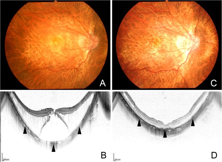
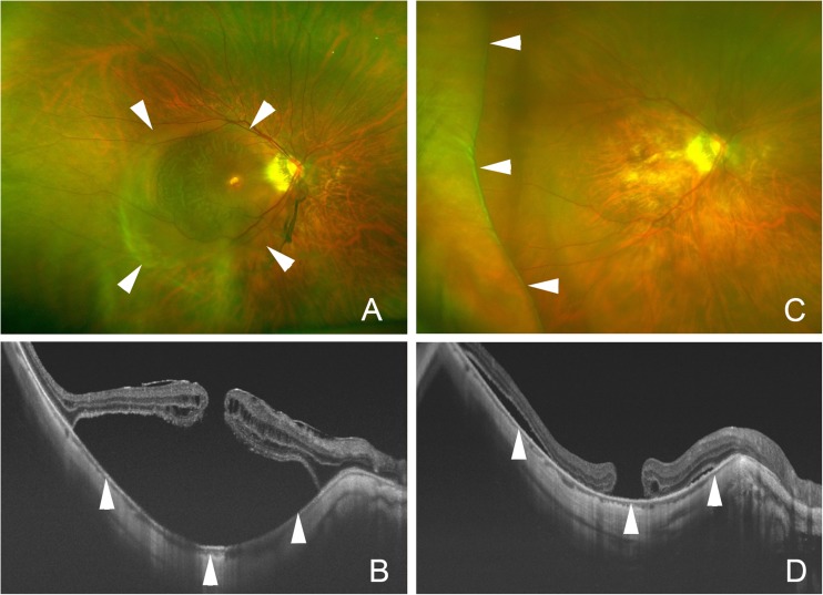
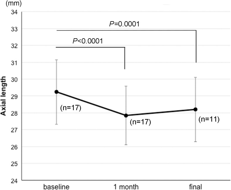
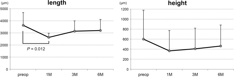
Results: The postoperative BCVA improved significantly from 0.76 ± 0.39 logarithm of the minimum angle of resolution (logMAR) units to 0.53 ± 0.35 logMAR units (P = 0.0004). The axial length decreased from 29.42 ± 1.81 mm to 27.97 ± 1.71 mm at 1 month. The MTM was resolved or decreased in all eyes. The MH was closed in 44 % of the MHRD eyes, and the retina was reattached in all of the MHRD eyes. The horizontal distance between the optic disc and the bottom of the posterior staphyloma was significantly decreased at 1 month (P = 0.012) but not at later times. The 3D-MRI images showed a reduction in the distance between the bottom of the posterior staphyloma and the center of the eye (P = 0.029) and a flattening of the posterior staphyloma (P = 0.010).
Conclusions: Vitrectomy with ILM peeling and scleral imbrication may be helpful in treating MTM and MHRD by reducing the degree of curvature of the posterior staphyloma.
Keywords: Macular hole retinal detachment; Myopic traction maculopathy; Optical coherence tomography; Scleral imbrication; three-dimensional magnetic resonance imaging.
Conflict of interest statement
Funding
No funding was received for this research.
Conflict of interest
All authors certify that they have no affiliations with or involvement in any organization or entity with any financial interest (such as honoraria; educational grants; participation in speakers’ bureaus; membership, employment, consultancies, stock ownership, or other equity interest; and expert testimony or patent-licensing arrangements), or non-financial interest (such as personal or professional relationships, affiliations, knowledge or beliefs) in the subject matter or materials discussed in this manuscript.
Ethical approval
All procedures performed in studies involving human participants were in accordance with the ethical standards of the institutional and/or national research committee and with the 1964 Helsinki Declaration and its later amendments or comparable ethical standards.
Informed consent
Informed consent was obtained from all individual participants included in the study.
Disclosure
The authors have no proprietary or commercial interest in any materials discussed in reporting these clinical observations and this article.
Figures
Similar articles
-
Scleral imbrication combined with vitrectomy and gas tamponade for refractory macular hole retinal detachment associated with high myopia.Retina. 2014 Dec;34(12):2451-7. doi: 10.1097/IAE.0000000000000246. Retina. 2014. PMID: 25062437
-
Vitrectomy with Internal Limiting Membrane Repositioning and Autologous Blood for Macular Hole Retinal Detachment in Highly Myopic Eyes.Ophthalmology. 2015 Sep;122(9):1889-98. doi: 10.1016/j.ophtha.2015.05.040. Epub 2015 Jul 2. Ophthalmology. 2015. PMID: 26143541
-
Long-term surgical outcomes of the inverted internal limiting membrane flap technique in highly myopic macular hole retinal detachment.Graefes Arch Clin Exp Ophthalmol. 2017 Jun;255(6):1101-1106. doi: 10.1007/s00417-017-3614-0. Epub 2017 Feb 20. Graefes Arch Clin Exp Ophthalmol. 2017. PMID: 28220252
-
Anatomical and visual outcomes in high myopic macular hole (HM-MH) without retinal detachment: a review.Graefes Arch Clin Exp Ophthalmol. 2014 Feb;252(2):191-9. doi: 10.1007/s00417-013-2555-5. Epub 2014 Jan 3. Graefes Arch Clin Exp Ophthalmol. 2014. PMID: 24384802 Review.
-
A meta-analysis of vitrectomy with or without internal limiting membrane peeling for macular hole retinal detachment in the highly myopic eyes.BMC Ophthalmol. 2016 Jun 13;16:87. doi: 10.1186/s12886-016-0266-5. BMC Ophthalmol. 2016. PMID: 27296383 Free PMC article. Review.
Cited by
-
Surgical classification for large macular hole: based on different surgical techniques results: the CLOSE study group.Int J Retina Vitreous. 2023 Jan 30;9(1):4. doi: 10.1186/s40942-022-00439-4. Int J Retina Vitreous. 2023. PMID: 36717928 Free PMC article.
-
Posterior pole retinotomy for treatment of recurrent macular hole retinal detachment in highly myopic eyes: a pilot study.BMC Ophthalmol. 2021 May 17;21(1):217. doi: 10.1186/s12886-021-01973-9. BMC Ophthalmol. 2021. PMID: 34001054 Free PMC article.
-
Lamellar macular hole in highly myopic eyes and insights into its development, evolution, and treatment: a mini-review.Graefes Arch Clin Exp Ophthalmol. 2024 Aug;262(8):2713-2724. doi: 10.1007/s00417-024-06419-8. Epub 2024 Feb 26. Graefes Arch Clin Exp Ophthalmol. 2024. PMID: 38407591 Review.
-
The Role of Internal Limiting Membrane Flap for Highly Myopic Macular Hole Retinal Detachment: Improving the Closure Rate but Leading to Excessive Gliosis.Front Med (Lausanne). 2021 Dec 23;8:812693. doi: 10.3389/fmed.2021.812693. eCollection 2021. Front Med (Lausanne). 2021. PMID: 35004792 Free PMC article.
-
Combined rhegmatogenous and tractional retinal detachment in a child with incontinentia pigmenti managed by scleral imbrication with scleral buckle.BMJ Case Rep. 2023 Feb 14;16(2):e253738. doi: 10.1136/bcr-2022-253738. BMJ Case Rep. 2023. PMID: 36787928 Free PMC article.
References
-
- Hayashi K, Ohno-Matsui K, Shimada N, Moriyama M, Kojima A, Hayashi W, Yasuzumi K, Nagaoka N, Saka N, Yoshida T, Tokoro T, Mochizuki M. Long-term pattern of progression of myopic maculopathy: a natural history study. Ophthalmology. 2010;117:1595–1611. doi: 10.1016/j.ophtha.2009.11.003. - DOI - PubMed
MeSH terms
LinkOut - more resources
Full Text Sources
Other Literature Sources
Medical
