Review
. 2017 Jan;255(1):1-6.
doi: 10.1007/s00417-016-3548-y. Epub 2016 Nov 10.

The neurovascular unit and the pathophysiologic basis of diabetic retinopathy

Affiliations
Review

The neurovascular unit and the pathophysiologic basis of diabetic retinopathy

Thomas W Gardner et al. Graefes Arch Clin Exp Ophthalmol. 2017 Jan.

Abstract

Purpose: To relate the concept of the retinal neurovascular unit and its alterations in diabetes to the pathophysiology of diabetic retinopathy.

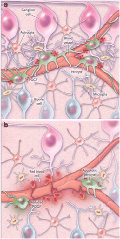
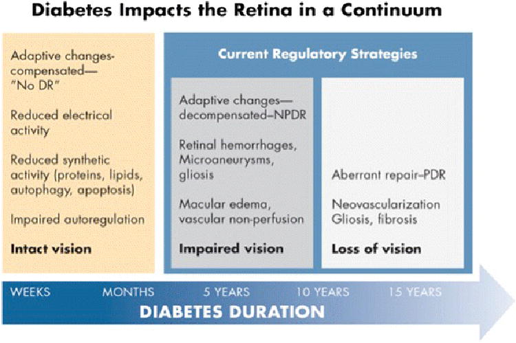
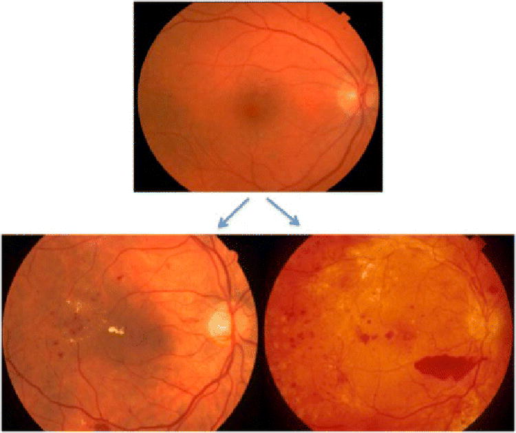
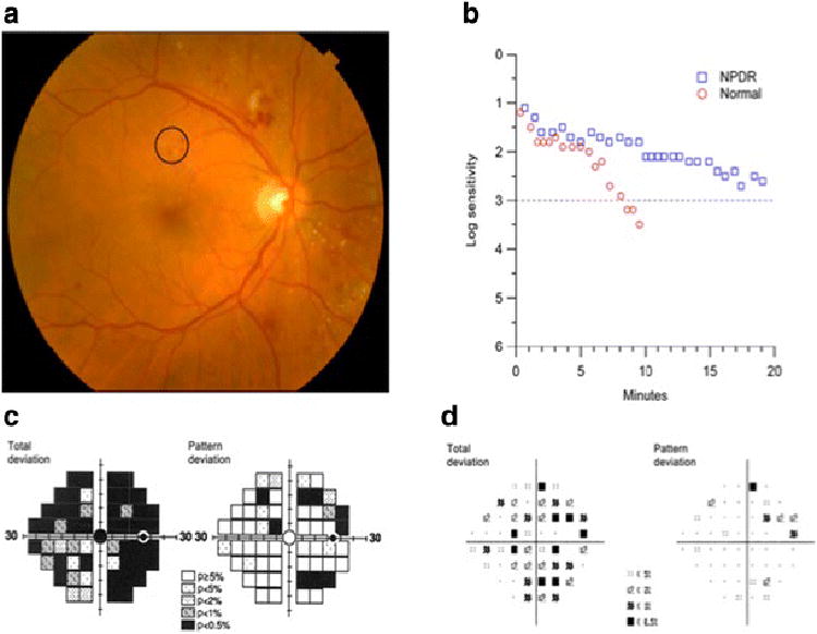
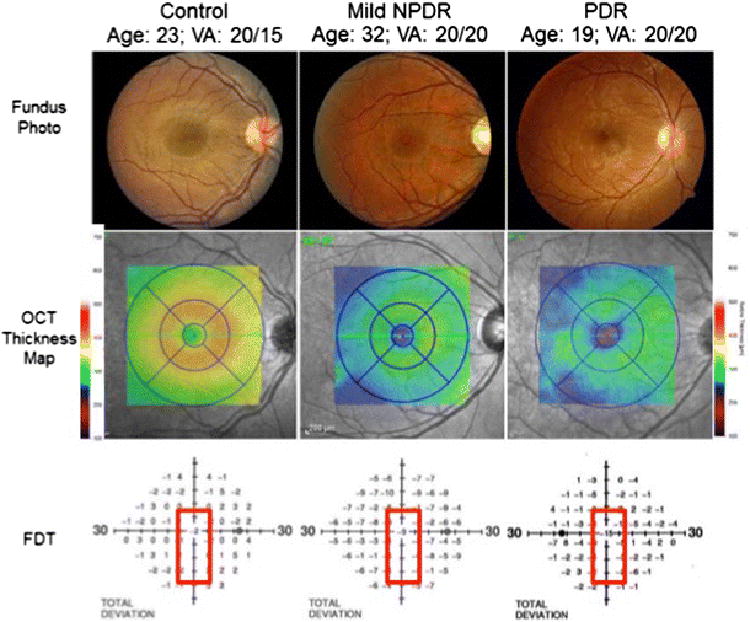
Methods: Case illustrations and conceptual frameworks are presented that illustrate adaptive and maladaptive "dis-integration" of the retinal neurovascular unit with the progression of diabetes.

Results: Retinopathy treatment should address pathophysiologic processes rather than pathologic lesions as is current practice.

Conclusions: Future improvements in the treatment of diabetic retinopathy requires deeper understanding of the cellular and molecular changes induced by diabetes, coupled with the use of quantitative phenotyping methods that assess the pathophysiologic processes.

Keywords: Diabetic retinopathy; Frequency-doubling perimetry; Neurovascular unit; Sensory neuropathy; Spectral domain optical coherence tomography (SD-OCT).


Figures

Figure 1
Figure 1
The neurovascular unit in the retina. Panel A shows pericytes and glial cells which support neuronal function and help form to the blood-retina barrier. Panel B shows an altered cellular environment with increased VEGF from glial cells, loss of platelet-derived growth factor (PDGF), and release of inflammatory cytokines leading to a breakdown of the blood-retina barrier and, in some cases, angiogenesis. From Antonetti (14), with permission.
Figure 2
Figure 2
A model of adaptive and maladaptive stages of diabetic retinopathy development. Within the first weeks to months of diabetes the retina adapts to a lower metabolic steady state with reduced electrical and biosynthetic activity, and adaptive autophagy and apoptosis, and impaired autoregulation. Vision remains intact and there is no clinical evidence of diabetic retinopathy. After 5 – 10 years of diabetes adaptive mechanisms begin to decompensate with the appearance of mild nonproliferative retinopathy, and early impairment of vision. Aberrant repair can ensue with the onset of proliferative retinopathy and loss of vision. Regulatory strategies are currently limited to the latter two stages. Artwork by David Murrel, MFA.
Figure 3
Figure 3
Progression of retinal failure in diabetes. Retinal failure occurs after long-term decompensated adaptive responses to metabolic dysfunction. The top image shows compensated diabetic retinopathy in a patient with mild non-proliferative diabetic retinopathy. The bottom images are examples of decompensated non-proliferative retinopathy with macular edema (lower left) and proliferative diabetic retinopathy with preretinal hemorrhage (lower right). From Gray (22) with permission.
Figure 4
Figure 4
A patient with 21 years of type 2 diabetes mellitus, 20/25 visual acuity and moderate nonproliferative diabetic retinopathy (A) exhibits severe visual function abnormalities. Dark adaptation (B) was severely delayed compared to normal adult responses. Frequency doubling perimetry fields (C) exhibited deep deficits in sensitivity that exceeded those revealed by standard photopic visual fields (D). From Jackson et al (34) with permission.
Figure 5
Figure 5
Color fundus photographs (top panel) of a healthy person (left), a person with mild non-proliferative diabetic retinopathy (NPDR) (middle), and a person with proliferative diabetic retinopathy (PDR) (right) demonstrate preserved macular anatomy without gross vascular compromise and visual acuities 20/20 or better. Spectral-domain optical coherence tomography (OCT) macular thickness mapping (central panel) demonstrates foveal and parafoveal thicknesses reduced by between 50 and 75 microns comparing healthy eyes to those with mild NPDR or PDR. The lower panel demonstrates a concomitant decline in foveal sensitivities in the diabetic patients as measured by frequency doubling perimetry, with a 9 decibel (dB) visual field sensitivity loss in the patient with mild NPDR and a 15 dB sensitivity loss in the patient with PDR.

References

    1. Hawkins BT, Davis TP. The blood-brain barrier/neurovascular unit in health and disease. Pharmacol Rev. 2005;57:173–185. - PubMed
    1. Metea MR, Newman EA. Signalling within the neurovascular unit in the mammalian retina. Exp Physiol. 2007;92:635–640. - PMC - PubMed
    1. Lott ME, Slocomb JE, Shivkumar V, Smith B, Gabbay RA, Quillen D, Gardner TW, Bettermann K. Comparison of retinal vasodilator and constrictor responses in type 2 diabetes. Acta ophthalmologica. 2012;90:e434–441. - PubMed
    1. Pemp B, Garhofer G, Weigert G, Karl K, Resch H, Wolzt M, Schmetterer L. Reduced retinal vessel response to flicker stimulation but not to exogenous nitric oxide in type 1 diabetes. Investigative ophthalmology & visual science. 2009;50:4029–4032. - PubMed
    1. Newman EA. Glial cell regulation of neuronal activity and blood flow in the retina by release of gliotransmitters. Philos Trans R Soc Lond B Biol Sci. 2015;370 - PMC - PubMed

MeSH terms