. 2017 Aug;37(6):1055-1066.
doi: 10.1007/s10571-016-0438-0. Epub 2016 Nov 10.

mitoLUHMES: An Engineered Neuronal Cell Line for the Analysis of the Motility of Mitochondria

Affiliations

mitoLUHMES: An Engineered Neuronal Cell Line for the Analysis of the Motility of Mitochondria

Tomasz M Stępkowski et al. Cell Mol Neurobiol. 2017 Aug.

Abstract

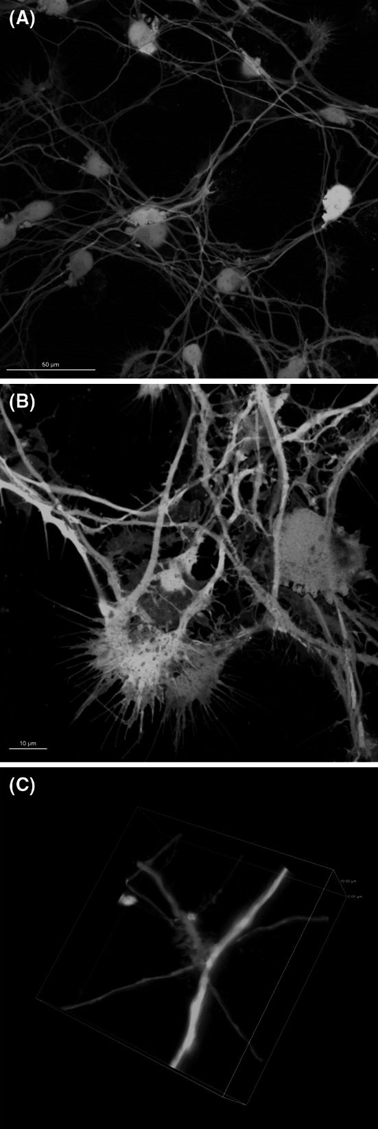
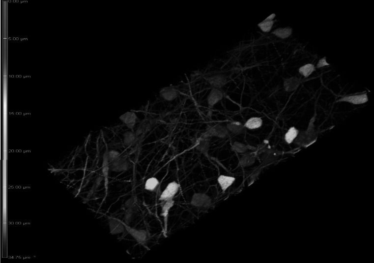
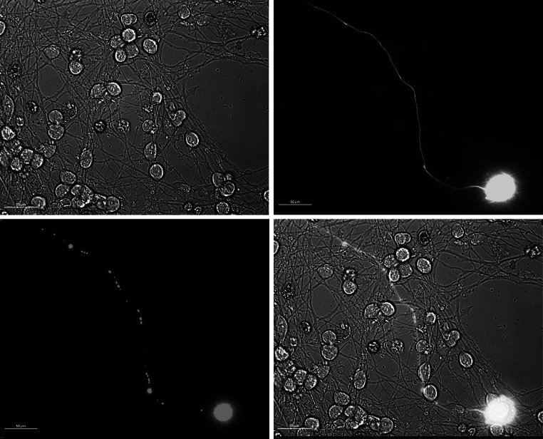
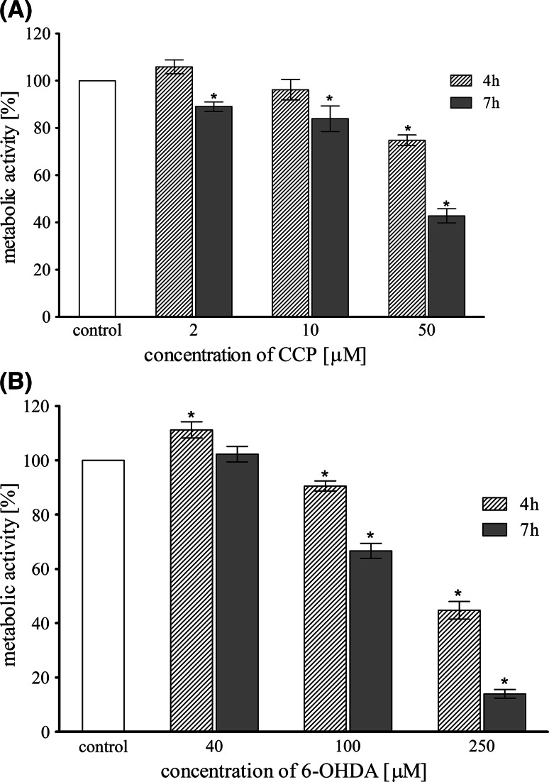
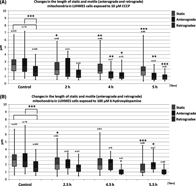
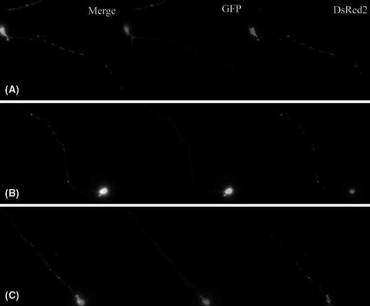
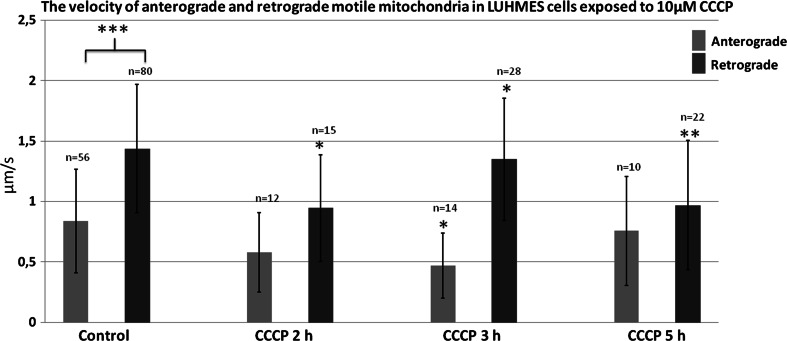
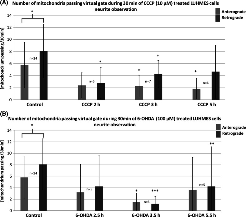
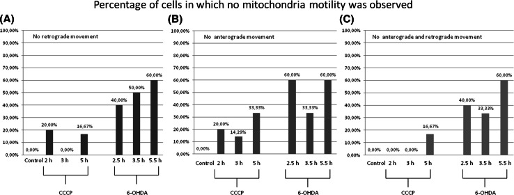
Perturbations in the transport of mitochondria and their quality control in neuronal cells underlie many types of neurological pathologies, whereas systems enabling convenient analysis of mitochondria behavior in cellular models of neurodegenerative diseases are limited. In this study, we present a modified version of lund human mesencephalic cells, mitoLUHMES, expressing GFP and mitochondrially targeted DsRed2 fluorescent proteins, intended for in vitro analysis of mitochondria trafficking by real-time fluorescence microscopy. This cell line can be easily differentiated into neuronal phenotype and allows us to observe movements of single mitochondria in single cells grown in high-density cultures. We quantified the perturbations in mitochondria morphology and dynamics in cells treated with model neurotoxins: carbonyl cyanide m-chlorophenylhydrazone and 6-hydroxydopamine. For the first time we filmed the processes of fission, fusion, pausing, and reversal of mitochondria movement direction in LUHMES cells. We present a detailed analysis of mitochondria length, velocity, and frequency of movement for static, anterograde, and retrograde motile mitochondria. The observed neurotoxin treatment-mediated decreases in morphological and kinetic parameters of mitochondria provide foundation for the future studies exploiting mitoLUHMES as a new model for neurobiology.

Keywords: 6-OHDA; CCCP; Live-cell imaging; Mitochondria motility; Neuronal models.


Conflict of interest statement

The authors declare that there is no personal or institutional conflict of interest related to the presented research and its publication.

Figures

Fig. 1
Fig. 1
The morphology of differentiated LUHMES cells. Confocal image of: a highly branched GFP-expressing LUHMES neuronal culture (magnification ×60). b Growth cones in GFP-expressing differentiated LUHMES cells. c Neurite branching and dendritic spikes in GFP-expressing differentiated LUHMES
Fig. 2
Fig. 2
The 3d reconstruction of the high-density neuronal network of differentiated GFP-expressing LUHMES cells mixed with wild-type LUHMES at proportions 1:20. The depth of the network reaches 30–35 μm. The depth color coding is displayed on the left border of the image
Fig. 3
Fig. 3
mitoDsRed2 and GFP-expressing mitoLUHMES cells mixed with wild-type LUHMES cells at proportion 1:200 allow visualization of individual mitochondria in a single neurite. The cells in this sample were treated with 6-OHDA for 5.5 h and therefore a fragmented circular mitochondria are clearly visible
Fig. 4
Fig. 4
Metabolic activity (resazurin assay) of LUHMES cells treated with different concentrations of CCCP (a) and 6-OHDA (b) for 4 and 7 h. Data are expressed as a percent of control, mean ± SD from three independent experiments (asterisk denotes statistically significant difference from vehicle-treated control, p < 0.05)
Fig. 5
Fig. 5
The length of static (green), anterograde (blue), and retrograde (red) mitochondria treated with CCCP (a) or 6-OHDA (b) in indicated time points. The results are presented as box-whiskers plots. Median values are shown as boxes borderline. The box top–bottom values are defined by the 25th and 75th percentile. The ends of the whiskers represent the minimum and maximum values. The n value represents the number of measured mitochondria. The statistical significance between the static, retrograde, and anterograde groups and corresponding controls is shown as asterisk. *p < 0.05. **p < 0.005; ***p < 0.0005
Fig. 6
Fig. 6
The morphology of mitochondria in mitoLUHMES cells grown with wt LUHMES at proportion of 1:200 a Vehicle-treated control cells (0.1% DMSO 5 h); b 5.5 h of incubation with 100 μM 6-OHDA; c 5 h of incubation with 10 μM CCCP
Fig. 7
Fig. 7
The mean velocity of anterograde and retrograde motile mitochondria in cells treated with CCCP for 2, 3 and 5 h. The error bars represent ±standard deviation. The statistical significance of the difference between retrograde mitochondria velocity in control and CCCP-treated cells is marked by asterisk: *p < 0.05. **p < 0.005; ***p < 0.0005
Fig. 8
Fig. 8
Mean frequency of events of transport of mitochondria in differentiated LUHMES cells treated with a CCCP and b 6-OHDA in indicated time points. The error bars represent ±standard deviation. The statistical significance between treated cells and control is denoted by asterisk *p < 0.05. **p < 0.005; ***p < 0.0005. The n indicates number of replicates (cells analyzed)
Fig. 9
Fig. 9
Percentage of mitoLUHMES cells in which mitochondria motility was stopped after treatment with indicated concentrations of CCCP and 6-OHDA. The left (red) panel—percentage of cells in which at least retrograde movement was not present; the inner panel (blue)—the percentage of cells in which at least anterograde movement was not present; the right panel (green)—the percentage of cells in which we did not observe any motile mitochondria

References

    1. Ashrafi G, Schlehe JS, LaVoie MJ, Schwarz TL (2014) Mitophagy of damaged mitochondria occurs locally in distal neuronal axons and requires PINK1 and Parkin. J Cell Biol 206:655–670 - PMC - PubMed
    1. Barnhart EL (2016) Mechanics of mitochondrial motility in neurons. Curr Opin Cell Biol 38:90–99 - PubMed
    1. Bertholet AM, Delerue T, Millet AM et al (2016) Mitochondrial fusion/fission dynamics in neurodegeneration and neuronal plasticity. Neurobiol Dis 90:3–19. doi:10.1016/j.nbd.2015.10.011 - PubMed
    1. Bhabha G, Johnson GT, Schroeder CM, Vale RD (2016) How dynein moves along microtubules. Trends Biochem Sci 41:94–105. doi:10.1016/j.tibs.2015.11.004 - PMC - PubMed
    1. Blesa J, Trigo-Damas I, Quiroga-Varela A, Jackson-Lewis VR (2015) Oxidative stress and Parkinson’s disease. Front Neuroanat 9:91. doi:10.3389/fnana.2015.00091 - PMC - PubMed

MeSH terms

LinkOut - more resources