

Diaphragm electromyographic activity following unilateral midcervical contusion injury in rats
- PMID: 27832610
- PMCID: PMC5288488
- DOI: 10.1152/jn.00727.2016
Diaphragm electromyographic activity following unilateral midcervical contusion injury in rats
Abstract
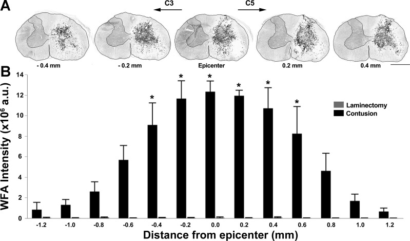
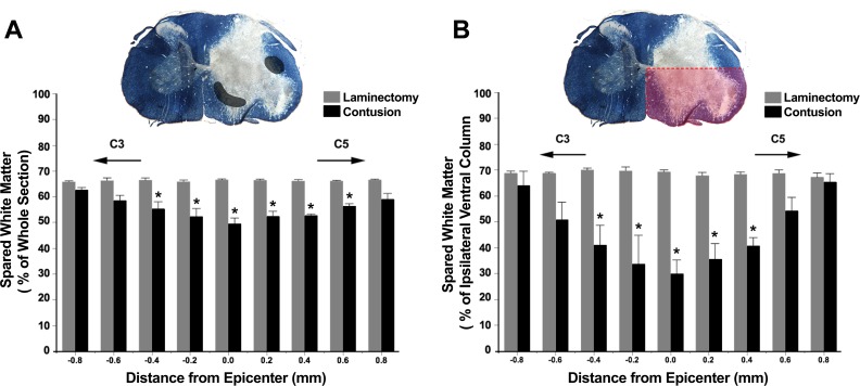
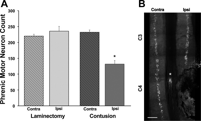
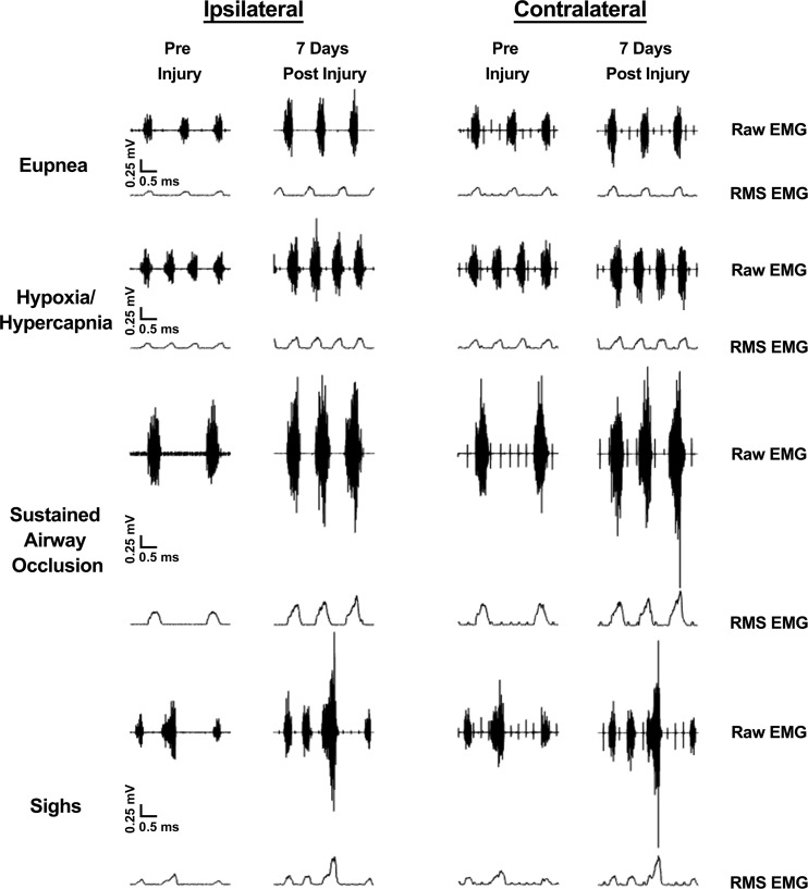
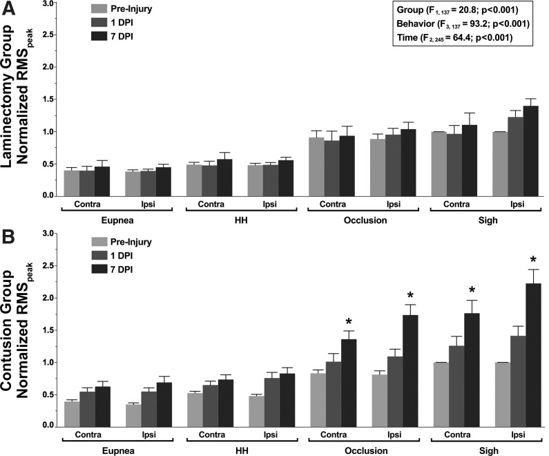
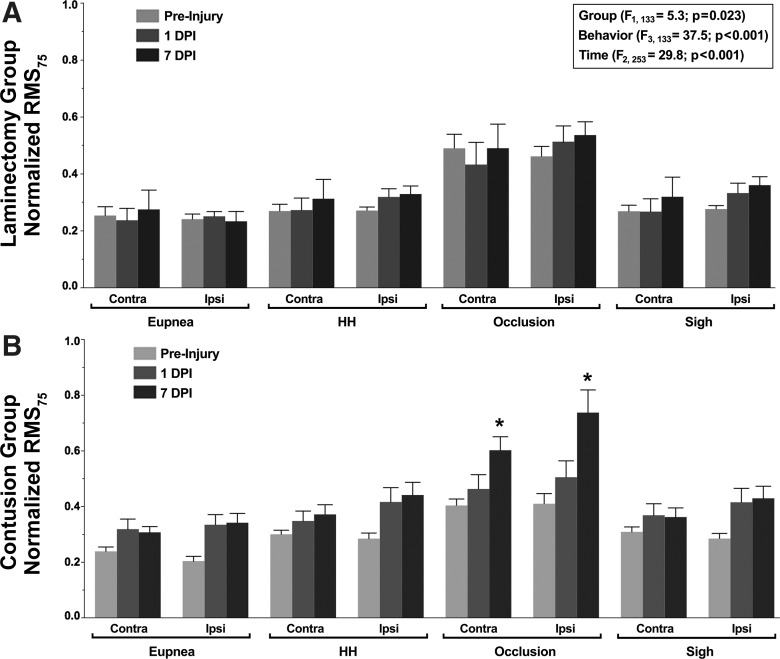
Contusion-type injuries to the spinal cord are characterized by tissue loss and disruption of spinal pathways. Midcervical spinal cord injuries impair the function of respiratory muscles and may contribute to significant respiratory complications. This study systematically assessed the impact of a 100-kDy unilateral C4 contusion injury on diaphragm muscle activity across a range of motor behaviors in rats. Chronic diaphragm electromyography (EMG) was recorded before injury and at 1 and 7 days postinjury (DPI). Histological analyses assessed the extent of perineuronal net formation, white-matter sparing, and phrenic motoneuron loss. At 7 DPI, ∼45% of phrenic motoneurons were lost ipsilaterally. Relative diaphragm root mean square (RMS) EMG activity increased bilaterally across a range of motor behaviors by 7 DPI. The increase in diaphragm RMS EMG activity was associated with an increase in neural drive (RMS value at 75 ms after the onset of diaphragm activity) and was more pronounced during higher force, nonventilatory motor behaviors. Animals in the contusion group displayed a transient decrease in respiratory rate and an increase in burst duration at 1 DPI. By 7 days, following midcervical contusion, there was significant perineuronal net formation and white-matter loss that spanned 1 mm around the injury epicenter. Taken together, these findings are consistent with increased recruitment of remaining motor units, including more fatigable, high-threshold motor units, during higher force, nonventilatory behaviors. Changes in diaphragm EMG activity following midcervical contusion injury reflect complex adaptations in neuromotor control that may increase the risk of motor-unit fatigue and compromise the ability to sustain higher force diaphragm efforts.
New & noteworthy: The present study shows that unilateral contusion injury at C4 results in substantial loss of phrenic motoneurons but increased diaphragm muscle activity across a range of ventilatory and higher force, nonventilatory behaviors. Measures of neural drive indicate increased descending input to phrenic motoneurons that was more pronounced during higher force, nonventilatory behaviors. These findings reveal novel, complex adaptations in neuromotor control following injury, suggestive of increased recruitment of more fatigable, high-threshold motor units.
Keywords: chronic EMG recordings; neuromotor control; respiratory muscles; spinal cord injury.
Copyright © 2017 the American Physiological Society.
Figures
Similar articles
-
The Impact of Midcervical Contusion Injury on Diaphragm Muscle Function.J Neurotrauma. 2016 Mar 1;33(5):500-9. doi: 10.1089/neu.2015.4054. Epub 2015 Nov 19. J Neurotrauma. 2016. PMID: 26413840 Free PMC article.
-
BDNF effects on functional recovery across motor behaviors after cervical spinal cord injury.J Neurophysiol. 2017 Feb 1;117(2):537-544. doi: 10.1152/jn.00654.2016. Epub 2016 Nov 9. J Neurophysiol. 2017. PMID: 27832605 Free PMC article.
-
Phrenic motor neuron degeneration compromises phrenic axonal circuitry and diaphragm activity in a unilateral cervical contusion model of spinal cord injury.Exp Neurol. 2012 Jun;235(2):539-52. doi: 10.1016/j.expneurol.2012.03.007. Epub 2012 Mar 23. Exp Neurol. 2012. PMID: 22465264
-
Descending bulbospinal pathways and recovery of respiratory motor function following spinal cord injury.Respir Physiol Neurobiol. 2009 Nov 30;169(2):115-22. doi: 10.1016/j.resp.2009.08.004. Epub 2009 Aug 12. Respir Physiol Neurobiol. 2009. PMID: 19682608 Review.
-
Spinal cord injury and diaphragm neuromotor control.Expert Rev Respir Med. 2020 May;14(5):453-464. doi: 10.1080/17476348.2020.1732822. Epub 2020 Feb 25. Expert Rev Respir Med. 2020. PMID: 32077350 Free PMC article. Review.
Cited by
-
Targeting drug or gene delivery to the phrenic motoneuron pool.J Neurophysiol. 2023 Jan 1;129(1):144-158. doi: 10.1152/jn.00432.2022. Epub 2022 Nov 23. J Neurophysiol. 2023. PMID: 36416447 Free PMC article. Review.
-
Quantifying mitochondrial volume density in phrenic motor neurons.J Neurosci Methods. 2021 Apr 1;353:109093. doi: 10.1016/j.jneumeth.2021.109093. Epub 2021 Feb 4. J Neurosci Methods. 2021. PMID: 33549636 Free PMC article.
-
Hypercapnia impacts neural drive and timing of diaphragm neuromotor control.J Neurophysiol. 2024 Dec 1;132(6):1966-1976. doi: 10.1152/jn.00466.2024. Epub 2024 Nov 16. J Neurophysiol. 2024. PMID: 39548981
-
A Simple, Low-Cost Implant for Reliable Diaphragm EMG Recordings in Awake, Behaving Rats.eNeuro. 2025 Feb 19;12(2):ENEURO.0444-24.2025. doi: 10.1523/ENEURO.0444-24.2025. Print 2025 Feb. eNeuro. 2025. PMID: 39890457 Free PMC article.
-
Mid-cervical spinal cord contusion causes robust deficits in respiratory parameters and pattern variability.Exp Neurol. 2018 Aug;306:122-131. doi: 10.1016/j.expneurol.2018.04.005. Epub 2018 Apr 10. Exp Neurol. 2018. PMID: 29653187 Free PMC article.
References
-
- Awad BI, Warren PM, Steinmetz MP, Alilain WJ. The role of the crossed phrenic pathway after cervical contusion injury and a new model to evaluate therapeutic interventions. Exp Neurol 248C: 398–405, 2013. - PubMed
-
- Bellingham MC. Synaptic inhibition of cat phrenic motoneurons by internal intercostal nerve stimulation. J Neurophysiol 82: 1224–1232, 1999. - PubMed
-
- Bellingham MC, Lipski J. Respiratory interneurons in the C-5 segment of the spinal cord of the cat. Brain Res 533: 141–146, 1990. - PubMed
Publication types
MeSH terms
Substances
Grants and funding
LinkOut - more resources
Full Text Sources
Other Literature Sources
Medical
Miscellaneous