

Calcitriol stimulates gene expression of cathelicidin antimicrobial peptide in breast cancer cells with different phenotype
- PMID: 27832772
- PMCID: PMC5103596
- DOI: 10.1186/s12929-016-0298-4
Calcitriol stimulates gene expression of cathelicidin antimicrobial peptide in breast cancer cells with different phenotype
Abstract
Background: In normal and neoplastic cells, growth-promoting, proangiogenic, cytotoxic and pro-apoptotic effects have all been attributed to cathelicidin antimicrobial peptide (CAMP). Nevertheless, little is known about the factors regulating this peptide expression in breast cancer. Herein we asked if the well-known antineoplastic hormone calcitriol could differentially modulate CAMP gene expression in human breast cancer cells depending on the cell phenotype in terms of efficacy and potency.
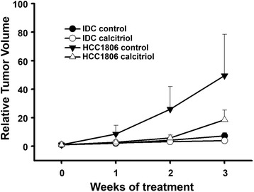
Methods: The established breast cancer cell lines MCF7, BT-474, HCC1806, HCC1937, SUM-229PE and a primary cell culture generated from invasive ductal breast carcinoma were used in this study. Calcitriol regulation of cathelicidin gene expression in vitro and in human breast cancer xenografts was studied by real time PCR. Tumorigenicity was evaluated for each cell line in athymic mice.
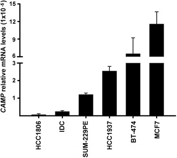
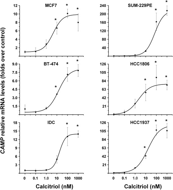
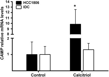
Results: Estrogen receptor (ER)α + breast cancer cells showed the highest basal CAMP gene expression. When incubated with calcitriol, CAMP gene expression was stimulated in a dose-dependent and cell phenotype-independent manner. Efficacy of calcitriol was lower in ERα + cells when compared to ERα- cells (<10 vs. >70 folds over control, respectively). Conversely, calcitriol lowest potency upon CAMP gene expression was observed in the ERα-/EGFR+ SUM-229PE cell line (EC50 = 70.8 nM), while the highest was in the basal-type/triple-negative cells HCC1806 (EC50 = 2.13 nM) followed by ERα + cells MCF7 and BT-474 (EC50 = 4.42 nM and 14.6 nM, respectively). In vivo, lower basal CAMP gene expression was related to increased tumorigenicity and lack of ERα expression. Xenografted triple-negative breast tumors of calcitriol-treated mice showed increased CAMP gene expression compared to vehicle-treated animals.
Conclusions: Independently of the cell phenotype, calcitriol provoked a concentration-dependent stimulation on CAMP gene expression, showing greater potency in the triple negative HCC1806 cell line. Efficacy of calcitriol was lower in ERα + cells when compared to ERα- cells in terms of stimulating CAMP gene expression. Lower basal CAMP and lack of ERα gene expression was related to increased tumorigenicity. Our results suggest that calcitriol anti-cancer therapy is more likely to induce higher levels of CAMP in ERα- breast cancer cells, when compared to ERα + breast cancer cells.
Keywords: Breast cancer; Calcitriol; Cathelicidin; LL-37; Vitamin D.
Figures
Similar articles
-
Calcitriol restores antiestrogen responsiveness in estrogen receptor negative breast cancer cells: a potential new therapeutic approach.BMC Cancer. 2014 Mar 29;14:230. doi: 10.1186/1471-2407-14-230. BMC Cancer. 2014. PMID: 24678876 Free PMC article.
-
The vitamin D analog EB1089 sensitizes triple-negative breast cancer cells to the antiproliferative effects of antiestrogens.Adv Med Sci. 2024 Sep;69(2):398-406. doi: 10.1016/j.advms.2024.08.004. Epub 2024 Sep 2. Adv Med Sci. 2024. PMID: 39233278
-
Activity of the antiestrogenic cajanin stilbene acid towards breast cancer.J Nutr Biochem. 2015 Nov;26(11):1273-82. doi: 10.1016/j.jnutbio.2015.06.004. Epub 2015 Jul 22. J Nutr Biochem. 2015. PMID: 26365581
-
Rosacea: The Blessing of the Celts - An Approach to Pathogenesis Through Translational Research.Acta Derm Venereol. 2016 Feb;96(2):147-56. doi: 10.2340/00015555-2220. Acta Derm Venereol. 2016. PMID: 26304030 Review.
-
The role of the basal stem cell of the human breast in normal development and cancer.Adv Exp Med Biol. 2011;720:121-34. doi: 10.1007/978-1-4614-0254-1_10. Adv Exp Med Biol. 2011. PMID: 21901623 Free PMC article. Review.
Cited by
-
Synergistic Antitumorigenic Activity of Calcitriol with Curcumin or Resveratrol is Mediated by Angiogenesis Inhibition in Triple Negative Breast Cancer Xenografts.Cancers (Basel). 2019 Nov 6;11(11):1739. doi: 10.3390/cancers11111739. Cancers (Basel). 2019. PMID: 31698751 Free PMC article.
-
Vitamin D and Its Role in Rheumatic Diseases.Metabolites. 2025 Apr 9;15(4):259. doi: 10.3390/metabo15040259. Metabolites. 2025. PMID: 40278388 Free PMC article. Review.
-
Calcitriol Inhibits the Proliferation of Triple-Negative Breast Cancer Cells through a Mechanism Involving the Proinflammatory Cytokines IL-1β and TNF-α.J Immunol Res. 2019 Apr 10;2019:6384278. doi: 10.1155/2019/6384278. eCollection 2019. J Immunol Res. 2019. PMID: 31093512 Free PMC article.
-
Transcriptomic profile induced by calcitriol in CaSki human cervical cancer cell line.PLoS One. 2025 Apr 1;20(4):e0319812. doi: 10.1371/journal.pone.0319812. eCollection 2025. PLoS One. 2025. PMID: 40168262 Free PMC article.
-
Identification of potential gene signatures associated with osteosarcoma by integrated bioinformatics analysis.PeerJ. 2021 May 27;9:e11496. doi: 10.7717/peerj.11496. eCollection 2021. PeerJ. 2021. PMID: 34123594 Free PMC article.
References
-
- Bruns H, Buttner M, Fabri M, Mougiakakos D, Bittenbring JT, Hoffmann MH, Beier F, Pasemann S, Jitschin R, Hofmann AD, et al. Vitamin D-dependent induction of cathelicidin in human macrophages results in cytotoxicity against high-grade B cell lymphoma. Sci Transl Med. 2015;7:282ra247. doi: 10.1126/scitranslmed.aaa3230. - DOI - PubMed
-
- Heilborn JD, Nilsson MF, Jimenez CI, Sandstedt B, Borregaard N, Tham E, Sorensen OE, Weber G, Stahle M. Antimicrobial protein hCAP18/LL-37 is highly expressed in breast cancer and is a putative growth factor for epithelial cells. Int J Cancer. 2005;114:713–719. doi: 10.1002/ijc.20795. - DOI - PubMed
MeSH terms
Substances
LinkOut - more resources
Full Text Sources
Other Literature Sources
Medical
Research Materials
Miscellaneous
