

Diagnosis of extraskeletal myxoid chondrosarcoma in the thigh using EWSR1-NR4A3 gene fusion: a case report
- PMID: 27832806
- PMCID: PMC5105269
- DOI: 10.1186/s13256-016-1113-2
Diagnosis of extraskeletal myxoid chondrosarcoma in the thigh using EWSR1-NR4A3 gene fusion: a case report
Abstract
Background: Extraskeletal myxoid chondrosarcoma is a rare soft tissue sarcoma that has unusual ultrastructural and molecular features. However, unlike other soft tissue sarcomas, it does not have specific clinical symptoms or radiological features, which can make its diagnosis difficult. Nevertheless, extraskeletal myxoid chondrosarcoma has a rare gene fusion (EWSR1-NR4A3) that is useful for making a differential diagnosis.
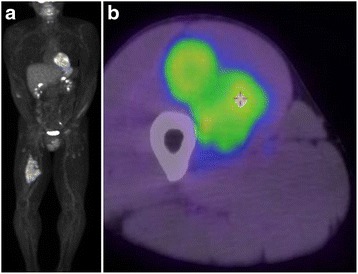
Case presentation: A 43-year-old Japanese man presented with a soft tissue mass in his right thigh. A physical examination and radiography revealed a large soft tissue mass. During magnetic resonance imaging, the mass exhibited isointensity on T1-weighted images and high intensity on T2-weighted images, as well as gadolinium enhancement at the side edge of the partition structure. Thus, we considered a possible diagnosis of a malignant myxoid soft tissue tumor, such as myxoid liposarcoma, myxofibrosarcoma, or metastatic carcinomas, including myoepithelial tumor and neuroendocrine tumor, and performed an incisional biopsy to make a definitive diagnosis. The pathological findings revealed a lobulated tumor with a myxoid structure and atypical spindle-shaped cells that created eosinophilic cord-like forms. Immunohistochemistry revealed that the tumor was positive for S-100 and negative for synaptophysin, chromogranin A, and pan keratin (AE1/AE3). The percentage of Ki-67 was 10 % in the hot spot area. Based on these clinicopathological findings, we initially considered the possibility of a myxoid liposarcoma, although we did not observe any lipoblasts. Therefore, we considered the possibility of an extraskeletal myxoid chondrosarcoma. As this tumor is very rare, we searched for the EWSR1-NR4A3 gene fusion using fluorescence in situ hybridization, which confirmed the diagnosis of extraskeletal myxoid chondrosarcoma. Positron emission tomography-computed tomography did not identify any obvious metastases, and we performed radical resection of our patient's vastus medialis and femur with a 3 cm margin. After the resection, we treated his resected femur using liquid nitrogen, and reconstructed his femur using autogenous fibula and plate fixation. No local recurrence or metastasis was observed at the 1-year follow-up.
Conclusion: Genetic testing is useful for diagnosing extraskeletal myxoid chondrosarcoma based on the presence of the EWSR1-NR4A3 gene fusion.
Keywords: EWSR1-NR4A3; Extraskeletal myxoid chondrosarcoma; Fluorescence in situ hybridization.
Figures


References
-
- Clark J, Benjamin H, Gill S, Sidhar S, Goodwin G, Crew J, et al. Fusion of the EWS gene to CHN, a member of the steroid/thyroid receptor gene superfamily, in a human myxoid chondrosarcoma. Oncogene. 1996;12:229–35. - PubMed
Publication types
MeSH terms
Substances
Supplementary concepts
LinkOut - more resources
Full Text Sources
Other Literature Sources
Medical
Research Materials
Miscellaneous
