. 2016 Nov 11:6:36803.
doi: 10.1038/srep36803.

Molecular inotropy mediated by cardiac miR-based PDE4D/PRKAR1α/phosphoprotein signaling

Affiliations

Molecular inotropy mediated by cardiac miR-based PDE4D/PRKAR1α/phosphoprotein signaling

Fikru B Bedada et al. Sci Rep. .

Abstract

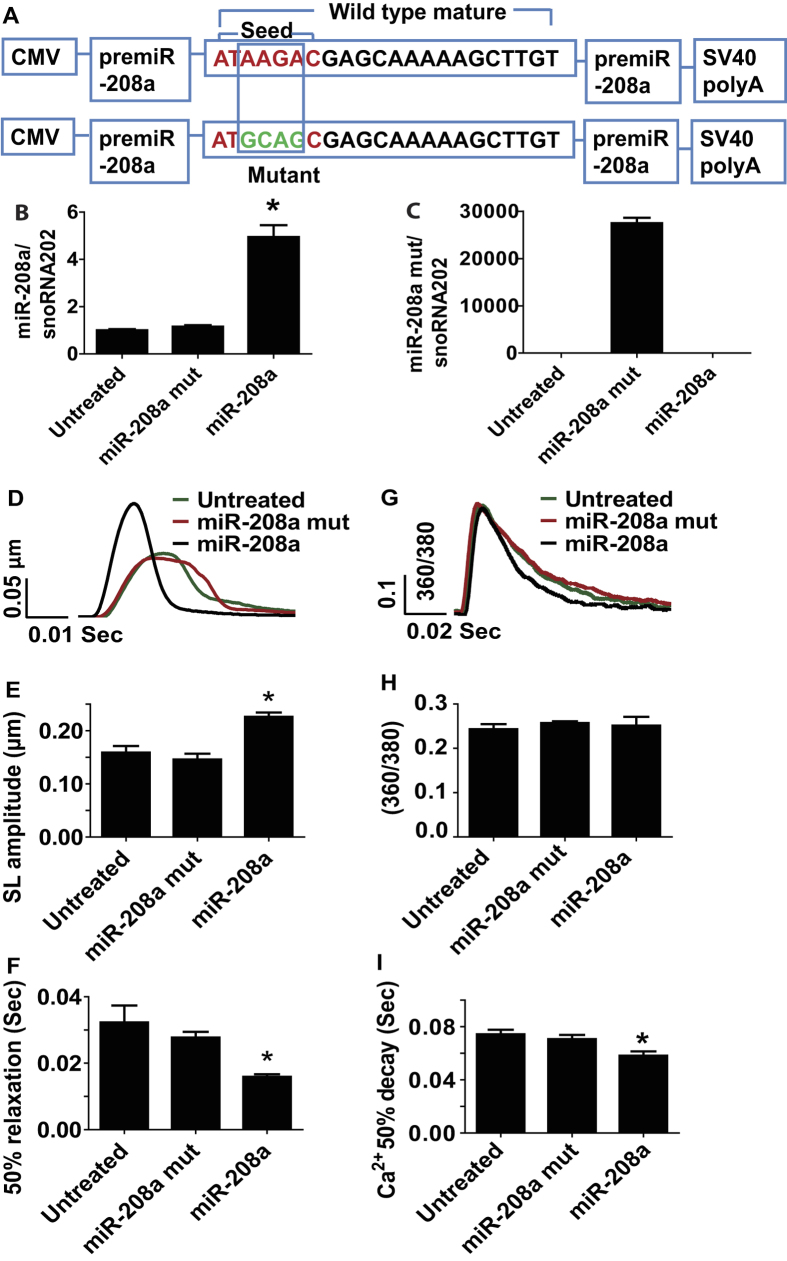
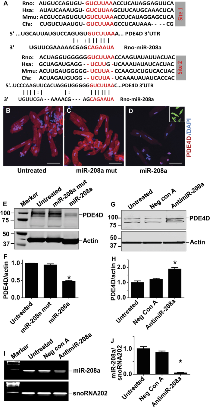
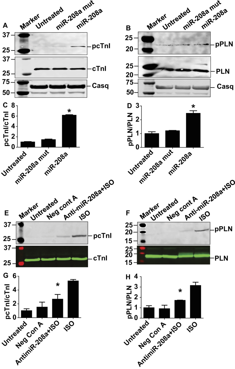
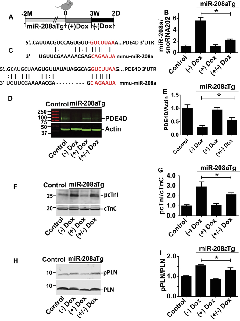
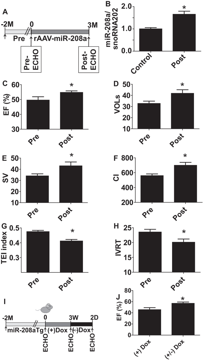
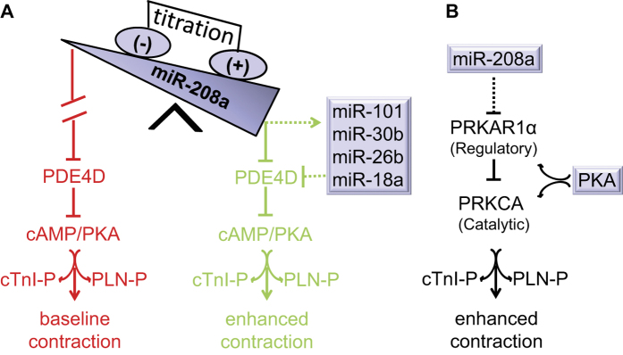
Molecular inotropy refers to cardiac contractility that can be modified to affect overall heart pump performance. Here we show evidence of a new molecular pathway for positive inotropy by a cardiac-restricted microRNA (miR). We report enhanced cardiac myocyte performance by acute titration of cardiac myosin-embedded miR-208a. The observed positive effect was independent of host gene myosin effects with evidence of negative regulation of cAMP-specific 3',5'-cyclic phosphodiesterase 4D (PDE4D) and the regulatory subunit of PKA (PRKAR1α) content culminating in PKA-site dependent phosphorylation of cardiac troponin I (cTnI) and phospholamban (PLN). Further, acute inhibition of miR-208a in adult myocytes in vitro increased PDE4D expression causing reduced isoproterenol-mediated phosphorylation of cTnI and PLN. Next, rAAV-mediated miR-208a gene delivery enhanced heart contractility and relaxation parameters in vivo. Finally, acute inducible increases in cardiac miR-208a in vivo reduced PDE4D and PRKAR1α, with evidence of increased content of several complementary miRs harboring the PDE4D recognition sequence. Physiologically, this resulted in significant cardiac cTnI and PLN phosphorylation and improved heart performance in vivo. As phosphorylation of cTnI and PLN is critical to myocyte function, titration of miR-208a represents a potential new mechanism to enhance myocardial performance via the PDE4D/PRKAR1α/PKA phosphoprotein signaling pathway.


Figures

Figure 1
Figure 1. MiR-208a confers positive inotropy and lusitropy to adult ventricular myocytes.
Constructs used for the generation of adenovirus expressing miR-208a (AdmiR-208a) and miR-208a mutant in adult myocytes (A). Expression of miR-208a and miR-208a mutant in cardiac myocytes (B,C). Superimposed representative SL shortening traces collected from field stimulated (0.5 Hz) untreated control (green), miR-208a mutant (red) and miR-208a-transduced (black) cardiac myocytes after gene transfer (D). Summaries of cardiac contraction and relaxation parameters, including SL shortening amplitude (E) and time to base line 50% (F). Data are shown as the means+/− SEM n = 27–31 myocytes. *P < 0.05 by One-way ANOVA. Representative calcium traces collected from field stimulated (0.5 Hz) untreated control (green), miR-208a mutant (red) and miR-208a-transduced (black) myocytes after gene transfer (G). Summaries of calcium amplitude and calcium decay parameters, including calcium amplitude (H) and 50% decay rate (I). Data are shown as mean +/− SEM, n = 9–11 myocytes. *P < 0.05 by One-way ANOVA.
Figure 2
Figure 2. MiR-208a suppresses PDE4D in adult ventricular myocytes.
(A) Two sites of PDE4D 3′ UTR sequence alignment are shown across species highlighting the seed match sequences of PDE4D 3′UTR (red) complementary to the seed sequences of miR-208a. Expression of PDE4D as assessed by immunohistochemistry (B–D). Significant reduction of total PDE4D staining was observed using a PDE4D specific antibody both in the cytoplasm and nucleus of miR-208a transduced but not in control myocytes (B–D). Sarcomeric actinin (green) shows the presence of the myocytes that were also stained by PDE4D (D; small box green for actinin). The images were taken with 40x objective confocal microscopy and bar shows scale of 50 μm. Western blots show that PDE4D was reduced significantly in miR-208a transduced but not in untreated or miR-208a mutant myocytes (E,F). Effects of pharmacologic inhibition of miR-208a via miR-208a power inhibitor that increased PDE4D content (G,H). Note that the Western blot in G was run longer resulting in distinct band separation versus E. Acute suppression of miR-208a in adult ventricular cardiac myocytes by antimiR-208a (I,J). Data are shown as mean +/− SEM, n = 3 *P < 0.05.
Figure 3
Figure 3. MiR-208a increases phosphorylation of cTnI and PLN in adult cardiac myocytes.
Western blot analysis of key phosphoproteins regulating SR calcium handling, relaxation and contractility after miR-208a transduction. Here, an increase in phosphorylation of cTnI (PKA sites Ser23/24) (A,C) and increase in phosphorylation of PLN (PKA site Ser16) (B,D) as compared to untreated control and miR-208a mutant was observed. MiR-208a did not affect total content of cTnI and PLN (A,B). Calsequestrin was not altered with either miR-208a or miR-208a mutant transduction or non-transduced control myocytes (A,B) and is used as loading control and for quantification of pcTnI and pPLN in each sample. Acute suppression of miR208a expression by anti-miR-208a reduced ISO-induced phosphorylation of cTnI and PLN (E–H). Data are shown as mean +/− SEM, n = 3–5 *P < 0.05.
Figure 4
Figure 4. In vivo titration of miR-208a increases cardiac phospho-substrates by suppressing PDE4D.
Experimental timeline for transgenic mice (A) and summary of titration of miR-208a levels in vivo (B). Two sites showing alignment highlighting the seed match sequences of mouse PDE4D 3′UTR (red) that is complementary for the seed sequences of miR-208a (C). The PDE4D content (D,E), the phosphoprotein content for cTnI (F,G) and PLN (H,I) are shown with representative Westerns and summary plots. Data are shown as the means +/− SEM. N = 3 *P < 0.05. Abbreviations, (+) Dox = dox treated, (−) Dox = no dox, (+/−) Dox = dox treated for 3 weeks and dox removed for 2 days.
Figure 5
Figure 5. MiR-208a enhances contractility and relaxation function in vivo.
Experimental timeline for rAAV-miR-208a delivery (A) and summaries of miR-208a expression (B) and echocardiography-based EF (C), VOLs (D), stroke volume (E), Cardiac index (F), TEI index (G) and IVRT (H). Data are shown as mean +/− SEM, n = 13–14 mice; *P < 0.05 compared with the pretreatment group. Experimental timeline for in vivo titration of miR-208a in transgenic mice (I) and summary of echocardiography-based cardiac function following miR-208a titration (J). Data are shown as the mean +/− SEM, n = 27 (J); *P < 0.05.
Figure 6
Figure 6. Working model: miR-208a titration and contractile function.
Cardiac expression of miR208a suppresses PDE4D through engagement of the seed sequence of miR-208a to the seed-match sequence of PDE4D 3′ UTR. MiR-208a also up-regulates other miRs targeting PDE4D. Suppression of PDE4D by miR-208a and other miRs promotes increased phosphorylation of cTnI and PLN thereby enhancing cardiac function (A, Green). Baseline contractility is depicted in red (A). Suppression of regulatory subunit of PKA (PRKAR1α) by miR-208a leads to active catalytic subunit (PRKCA) and PKA activation (B). This concerted amplification of PKA signaling in turn increases phosphorylation of cTnI and PLN thereby enhancing cardiac function (A, green and B). The dotted line indicates indirect regulation.

References

    1. Francis G. S. Pathophysiology of chronic heart failure. Am J Med 110 Suppl 7A, 37S–46S (2001). - PubMed
    1. Bristow M. R. Pharmacogenetic targeting of drugs for heart failure. Pharmacology & therapeutics 134, 107–115, doi: 10.1016/j.pharmthera.01.002 (2012). - DOI - PubMed
    1. Hasenfuss G. & Teerlink J. R. Cardiac inotropes: current agents and future directions. European heart journal 32, 1838–1845, doi: 10.1093/eurheartj/ehr026 (2011). - DOI - PubMed
    1. Francis G. S., Bartos J. A. & Adatya S. Inotropes. J Am Coll Cardiol, 63(20), 2069–2078, doi: 10.1016/j.jacc.01.016 (2014). - DOI - PubMed
    1. Kiriakidou M. et al.. An mRNA m7G cap binding-like motif within human Ago2 represses translation. Cell 129, 1141–1151, doi: S0092-8674(07)00653-8 (2007). - PubMed

Publication types

MeSH terms

LinkOut - more resources