. 2016 Oct 27:4:117.
doi: 10.3389/fcell.2016.00117. eCollection 2016.

Extracellular Microvesicle Production by Human Eosinophils Activated by "Inflammatory" Stimuli

Affiliations

Extracellular Microvesicle Production by Human Eosinophils Activated by "Inflammatory" Stimuli

Praveen Akuthota et al. Front Cell Dev Biol. .

Abstract

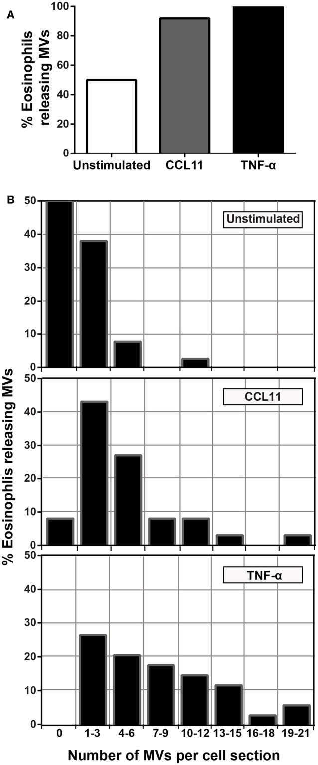
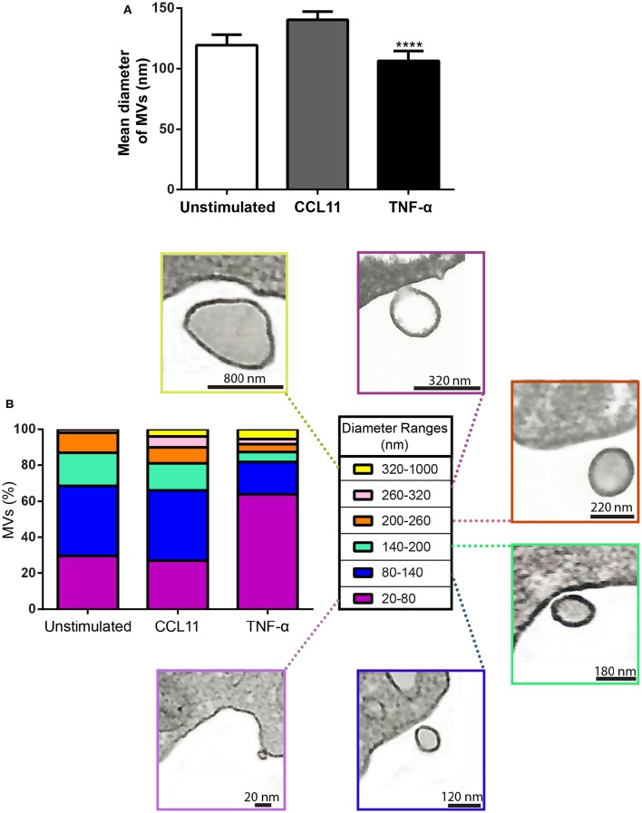
A key function of human eosinophils is to secrete cytokines, chemokines and cationic proteins, trafficking, and releasing these mediators for roles in inflammation and other immune responses. Eosinophil activation leads to secretion of pre-synthesized granule-stored mediators through different mechanisms, but the ability of eosinophils to secrete extracellular vesicles (EVs), very small vesicles with preserved membrane topology, is still poorly understood. In the present work, we sought to identify and characterize EVs released from human eosinophils during different conditions: after a culturing period or after isolation and stimulation with inflammatory stimuli, which are known to induce eosinophil activation and secretion: CCL11 (eotaxin-1) and tumor necrosis factor alpha (TNF-α). EV production was investigated by nanoscale flow cytometry, conventional transmission electron microscopy (TEM) and pre-embedding immunonanogold EM. The tetraspanins CD63 and CD9 were used as EV biomarkers for both flow cytometry and ultrastructural immunolabeling. Nanoscale flow cytometry showed that human eosinophils produce EVs in culture and that a population of EVs expressed detectable CD9, while CD63 was not consistently detected. When eosinophils were stimulated immediately after isolation and analyzed by TEM, EVs were clearly identified as microvesicles (MVs) outwardly budding off the plasma membrane. Both CCL11 and TNF-α induced significant increases of MVs compared to unstimulated cells. TNF-α induced amplified release of MVs more than CCL11. Eosinophil MV diameters varied from 20 to 1000 nm. Immunonanogold EM revealed clear immunolabeling for CD63 and CD9 on eosinophil MVs, although not all MVs were labeled. Altogether, we identified, for the first time, that human eosinophils secrete MVs and that this production increases in response to inflammatory stimuli. This is important to understand the complex secretory activities of eosinophils underlying immune responses. The contribution of the eosinophil-derived MVs to the regulation of immune responses awaits further investigation.

Keywords: CCL11 (eotaxin-1); CD63; CD9; cell secretion; inflammation; tetraspanins; transmission electron microscopy (TEM); tumor necrosis factor alpha (TNF-α).


Figures

Figure 1
Figure 1
TEM reveals production of MVs by human eosinophils. (A) A representative micrograph of a human eosinophil documents several MVs (highlighted in blue in Ai) budding directly from the plasma membrane. Eosinophil specific granules (Gr), the singular population of secretory granules in the cytoplasm, show lucencies in their granule cores, indicative of cell activation. Two lipid bodies (LB), which typically appear as very electron-dense organelles in eosinophils, are seen in the cell periphery. Eosinophils were isolated from the peripheral blood from healthy donors by negative selection, kept in medium during 1 h, immediately fixed while still in suspension and processed for conventional TEM. N, nucleus.
Figure 2
Figure 2
Identification of Eosinophil EVs by Nanoscale Flow Cytometry. (A) The ability of the AstriosEQ to discriminate sub-micrometer particles is shown using a mixture of Control Latex Beads (200, 300, and 500 nm). (B) Flow cytometry of RPMI with FBS (EV-depleted) alone showing non-specific events and electronic noise. Forward scatter and side scatter plotted. (C) Flow cytometry of human eosinophil EVs in RPMI with FBS (EV-depleted). Gate drawn around EV signal (FSC and SSC plotted). (D) Gated eosinophil EVs. (E) Human eosinophil EVs CD9 expression (blue) overlaid on IgG control (red). (F) Human eosinophil CD63 expression (blue) overlaid on IgG control (red) shows minimal detection of CD63 by nanoscale flow cytometry. Representative of four experiments from four individual donors. (G) Mean CD9 fluorescence (p = 0.13, paired t-test). Panels (B–G) are representative of four independent experiments from four individual human donors.
Figure 3
Figure 3
CCL11 and TNF-α stimulation induce release of MVs by human eosinophils. (A, Ai, B, Bi, Bii) MVs are seen at the surface of both unstimulated (A) and CCL11-stimulated (B) human eosinophils. Note in high magnification (Ai) that the phospholipid bilayer membrane, which is seen by TEM as a trilaminar structure (arrowheads), is observed around the EVs, plasma membrane and secretory granule (Gr) delimiting membrane. (Bii) Shows in high magnification a MV in final process of detaching from the plasma membrane (arrow). (C) Significant increases in numbers of MVs occurred after stimulation with CCL11 or TNF-α. Eosinophils were isolated from the peripheral blood by negative selection, stimulated for 1 h, immediately fixed and processed for conventional TEM. Counts were derived from three experiments with a total of 516 MVs counted in 110 electron micrographs randomly taken and showing the entire cell profile and nucleus (N). Data represent mean ± S.E.M. ***P < 0.002 (CCL11 vs. unstimulated); ****P < 0.0001 (TNF-α vs. unstimulated); ##P < 0.02 (TNF-α vs. CCL11).
Figure 4
Figure 4
Proportion of eosinophils releasing MVs. (A) While just 50% of unstimulated cells produced MVs, 90 and 100% of eosinophils formed MVs in CCL11 and TNF-α-stimulated eosinophils, respectively. (B) Heterogeneity of cell responses in unstimulated and stimulated eosinophils. In unstimulated cells, most MV-producing cells (30%) released 1–3 MVs/cell section whereas ~70% of cells produced 1–9 MVs and 4–21 MVs/cell section in response to CCL11 and TNF-α stimulation, respectively. Counts were derived from three experiments with a total of 516 MVs counted in 110 electron micrographs randomly taken and showing the entire cell profile and nucleus.
Figure 5
Figure 5
Differential release of nascent MVs by human activated eosinophils. (A) A representative electron micrograph of a TNF-α-stimulated eosinophil shows MVs in different steps of budding at cell surface (highlighted in blue, arrows) and secretory granules (Gr) exhibiting content losses in the cytoplasm. The MVs indicated by the black arrows are seen in high magnification in (Ai) and (Aii). Note, that while (Ai) shows a MV in process of outward budding; (Aii) shows a free MV, completely detached from the plasma membrane. (B) Illustration depicting the process of MV formation in human eosinophils as observed in the present work. (C) Significant increases in numbers of budding MVs occurred after stimulation with CCL11 or TNF-α compared to unstimulated cells (****P < 0.0001). TNF-α elicited higher numbers of MVs in process of budding compared to the CCL11 group (####P < 0.0001). Increase in numbers of free vesicles occurred after stimulation with CCL11 compared to unstimulated cells (*P = 0.020). Eosinophils were isolated from the peripheral blood by negative selection, stimulated for 1 h, immediately fixed in suspension and processed for conventional TEM. Counts were derived from three experiments, with a total of 516 MVs counted in 110 electron micrographs randomly taken and showing the entire cell profile and nucleus (N).
Figure 6
Figure 6
Diameter of MVs produced by unstimulated and stimulated human eosinophils. (A) Determination of mean diameter of MVs. Significant decrease in the diameters of released MVs occurred after stimulation with TNF-α compared to both unstimulated and CCL11-stimulated groups (****P < 0.0001). (B) The percentages of MVs per diameter ranges are shown. Representative electron micrographs of MVs are seen within each diameter range. Diameters of MVs were measured using Image J software and grouped in different ranges (20–100, 100–200, 200–300, 300–1000 nm). These analyses were done in clear cross-cell sections exhibiting the entire eosinophil cell profile (n = 110 cells), intact plasma membranes, and nuclei.
Figure 7
Figure 7
CD63 and CD9 immunolabeling of MVs by immunonanogold EM. (A–C) Representative electron micrographs of stimulated human eosinophils showing the entire cell profile after CD63 (A,C) or CD9 (B) immunolabeling. Note that while CD63 is consistently found intracellularly in association with secretory granules (Gr) as seen in (A) and (C), pools of CD9 are more detectable at the eosinophil surface as observed in (B). (Ai, Aii, Bi, Bii, Ci, Cii) CD63 and CD9-positive MVs (arrows) are seen in higher magnification in the boxed areas. Note, that not all MVs were labeled. Eosinophils were isolated from the peripheral blood by negative selection, stimulated for 1 h with CCL11 (A,B) or TNF-α (C), immediately fixed in suspension and processed for immunonanogold EM.
Figure 8
Figure 8
Annexin-V staining of human eosinophils. (A–C) Flow cytometric analyses show higher annexin-V intensities in CCL11 and TNF-α-stimulated compared to unstimulated cells. (D) Representative confocal microscopic image from a CCL11-stimulated eosinophil reveal cell surface distribution of annexin-V. Arrows indicate suggestive images of MV formation. Eosinophils were isolated from the peripheral blood by negative selection, stimulated for 1 h with CCL11 or TNF-α and stained with annexin-V.

Similar articles

Cited by

References

    1. Akuthota P., Capron K., Weller P. F. (2014). Eosinophil purification from peripheral blood. Methods Mol. Biol. 1178, 13–20. 10.1007/978-1-4939-1016-8_2 - DOI - PMC - PubMed
    1. Akuthota P., Melo R. C. N., Spencer L. A., Weller P. F. (2012). MHC Class II and CD9 in human eosinophils localize to detergent-resistant membrane microdomains. Am. J. Respir. Cell Mol. Biol. 46, 188–195. 10.1165/rcmb.2010-0335OC - DOI - PMC - PubMed
    1. Andreu Z., Yáñez-Mó M. (2014). Tetraspanins in extracellular vesicle formation and function. Front. Immunol. 5:442. 10.3389/fimmu.2014.00442 - DOI - PMC - PubMed
    1. Bandeira-Melo C., Gillard G., Ghiran I., Weller P. F. (2000). EliCell: a gel-phase dual antibody capture and detection assay to measure cytokine release from eosinophils. J. Immunol. Methods 244, 105–115. 10.1016/S0022-1759(00)00264-7 - DOI - PubMed
    1. Bandeira-Melo C., Perez S. A., Melo R. C. N., Ghiran I., Weller P. F. (2003). EliCell assay for the detection of released cytokines from eosinophils. J. Immunol. Methods 276, 227–237. 10.1016/S0022-1759(03)00076-0 - DOI - PubMed

LinkOut - more resources