. 2016 Nov 22;113(47):13378-13383.
doi: 10.1073/pnas.1616627113. Epub 2016 Nov 9.

Transcriptional start site heterogeneity modulates the structure and function of the HIV-1 genome

Affiliations

Transcriptional start site heterogeneity modulates the structure and function of the HIV-1 genome

Siarhei Kharytonchyk et al. Proc Natl Acad Sci U S A. .

Abstract

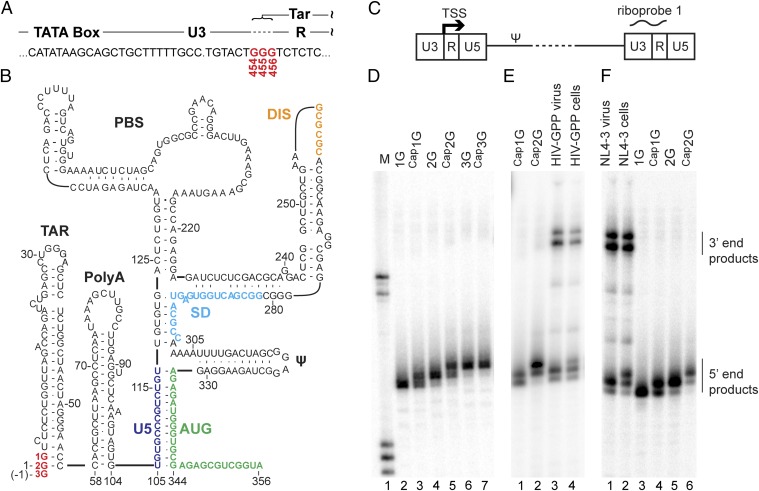
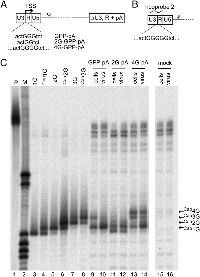
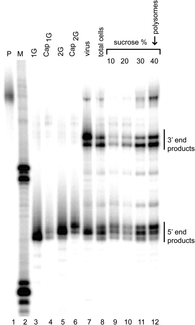
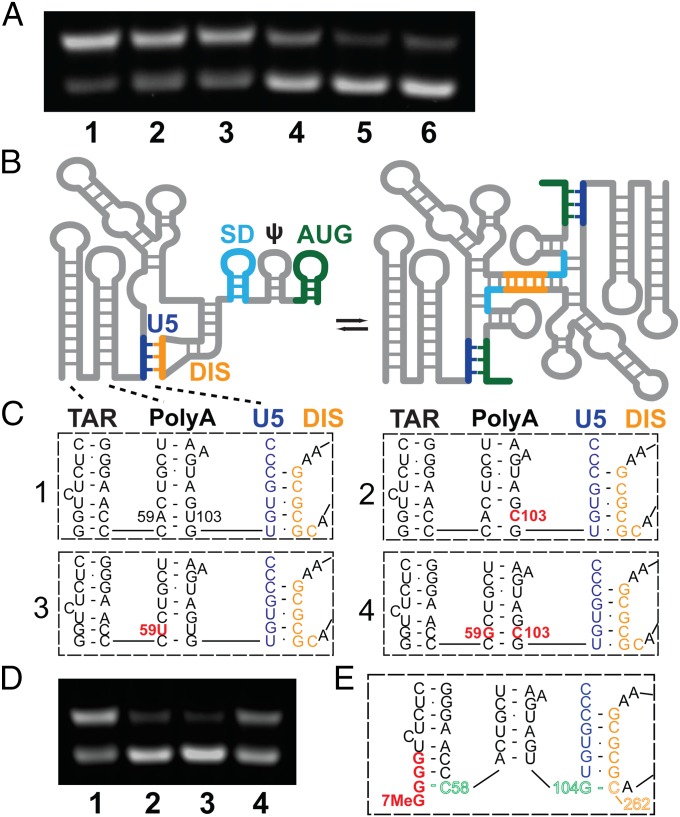
The promoter in HIV type 1 (HIV-1) proviral DNA contains three sequential guanosines at the U3-R boundary that have been proposed to function as sites for transcription initiation. Here we show that all three sites are used in cells infected with HIV-1 and that viral RNAs containing a single 5' capped guanosine (Cap1G) are specifically selected for packaging in virions, consistent with a recent report [Masuda et al. (2015) Sci Rep 5:17680]. In addition, we now show that transcripts that begin with two or three capped guanosines (Cap2G or Cap3G) are enriched on polysomes, indicating that RNAs synthesized from different transcription start sites have different functions in viral replication. Because genomes are selected for packaging as dimers, we examined the in vitro monomer-dimer equilibrium properties of Cap1G, Cap2G, and Cap3G 5'-leader RNAs in the NL4-3 strain of HIV-1. Strikingly, under physiological-like ionic conditions in which the Cap1G 5'-leader RNA adopts a dimeric structure, the Cap2G and Cap3G 5'-leader RNAs exist predominantly as monomers. Mutagenesis studies designed to probe for base-pairing interactions suggest that the additional guanosines of the 2G and 3G RNAs remodel the base of the PolyA hairpin, resulting in enhanced sequestration of dimer-promoting residues and stabilization of the monomer. Our studies suggest a mechanism through which the structure, function, and fate of the viral genome can be modulated by the transcriptionally controlled presence or absence of a single 5' guanosine.

Keywords: 5′-leader; HIV-1; RNA; structure; transcription.


Conflict of interest statement

The authors declare no conflict of interest.

Figures

Fig. 1.
Fig. 1.
HIV-1 RNA 5′-end heterogeneity in virions and cells. (A) The HIV-1NL4-3 provirus U3–R boundary showing the three guanosines (G454, G455, and G456) that can serve as the TSS (and encode the 5′ end of the TAR element of the viral transcript). (B) Secondary structure of the dimer-promoting form of HIV-1 RNA (adapted from ref. 42). 1G, 2G, and 3G guanosines (red) correspond to G456, G455, and G454 TSS residues. (C) Schematic representation of an HIV-1 provirus (not to scale). Ψ indicates the packaging signal. Riboprobe 1 indicates the location of the probe used in DF and Fig. 3. (D) RNase protection products of in vitro-generated HIV-1 5′-L RNA size standards corresponding to the use of alternate TSS, with or without capping. Lane 1: molecular size standards. Lanes 2–7: protected products for 1G, Cap1G, 2G, Cap2G, 3G, and Cap3G RNAs, respectively. (E) RNase protection analysis of cell and virus samples from transiently transfected cells. Lanes 1 and 2: protected fragments for Cap1G and Cap2G RNA standards. Lanes 3 and 4: fragments protected by virion and cell RNA, respectively. (F) RNase protection analysis of RNA samples harvested from CEM-SS cells chronically infected with HIV-1 strain NL4-3. Lanes 1 and 2: products protected by RNA samples from virus or cells, respectively. Lanes 3–6: protected fragments generated from the indicated RNA size standards. Mobilities of products protected from the 5′ and 3′ ends of viral RNA are indicated on the right.
Fig. 2.
Fig. 2.
Selectivity for packaging of Cap1G RNAs from cells with altered RNA proportions. (A) Schematic representation of constructs with altered numbers of guanosines at the U3/R junction used to skew intracellular RNA populations. ΔU3, R + pA indicates replacement of downstream LTR sequences with an SV40 polyadenylation signal. (B) Schematic representation of the portion of the HIV-1 genome to which riboprobe 2 is complementary. Note the five guanosines at the U3/R border, which allowed discrimination among more products than the 3G riboprobe. (C) RNase protection assay of constructs with altered numbers of guanosines. Lane 1: undigested probe. Lane 2: size standards. Lanes 3–8: fragments protected by RNA standards. Lanes 9–16: products protected by the indicated cell and virus RNA samples from cells transfected with the indicated HIV-1 GPP derivatives. Mobilities of products protected by Cap1G, Cap2G, Cap3G, and Cap4G RNAs are indicated at the right.
Fig. 3.
Fig. 3.
Analysis of HIV-1 RNA forms associated with polyribosomes. RNase protection assay on cell lysate fractions from chronically infected CEM-SS cells, analyzed with riboprobe 1 (Fig. 1). Lane 1: undigested probe. Lane 2: size standards. Lanes 3–6: fragments protected by the indicated RNA standards. Lanes 7–8: fragments protected by chronically infected cell medium (virus) and total cell lysate. Lanes 9–12: probe fragments protected by RNA from 10, 20, 30, and 40% sucrose step gradient fractions. Mobilities of protected products from the 5′ and 3′ ends of viral RNA are indicated on the right.
Fig. 4.
Fig. 4.
(A) Influence of 5′-guanosine number and capping on dimerization, as follows: lane 1: 1G 5′-L RNA; lane 2: Cap1G 5′-L; lane 3: 2G 5′-L; lane4: Cap2G 5′-L; lane 5: 3G 5′-L; and lane 6: Cap3G 5′-L. Under the conditions used, RNAs in lanes 1–3 favor the dimer, whereas those in lanes 4–6 favor the monomer. (BD) Mutations engineered to destabilize the base of PolyA favor the monomer. (B) Working model for secondary structural changes associated with the monomer–dimer equilibrium (adapted from ref. 26). (C) Proposed base pairing of residues in the lower stems of the TAR and PolyA hairpins and the U5:DIS pseudoknot for wild-type (1), U103C (2), A59U (3), and A59G/U103C (4) constructs. (D) Influence of point mutations on dimerization; lane numbers correspond to the panel labels in C. (E) Model for TSS-dependent dimerization control. In Cap3G transcripts, G(−1)–C58 base pairing disrupts and remodels the PolyA hairpin, allowing G104 to base pair with C262 of the DIS palindrome and thereby stabilizing the monomeric conformer.

Similar articles

Cited by

References

    1. Coffin JM, Hughes SH, Varmus HE. Retroviruses. Cold Spring Harbor Lab Press; Plainview, NY: 1997. - PubMed
    1. Chiu YL, Coronel E, Ho CK, Shuman S, Rana TM. HIV-1 Tat protein interacts with mammalian capping enzyme and stimulates capping of TAR RNA. J Biol Chem. 2001;276(16):12959–12966. - PubMed
    1. Zhou M, et al. The Tat/TAR-dependent phosphorylation of RNA polymerase II C-terminal domain stimulates cotranscriptional capping of HIV-1 mRNA. Proc Natl Acad Sci USA. 2003;100(22):12666–12671. - PMC - PubMed
    1. Menees TM, Müller B, Kräusslich HG. The major 5′ end of HIV type 1 RNA corresponds to G456. AIDS Res Hum Retroviruses. 2007;23(8):1042–1048. - PubMed
    1. Sharma A, Yilmaz A, Marsh K, Cochrane A, Boris-Lawrie K. Thriving under stress: Selective translation of HIV-1 structural protein mRNA during Vpr-mediated impairment of eIF4E translation activity. PLoS Pathog. 2012;8(3):e1002612. - PMC - PubMed

Publication types