. 2016 Nov 22;113(47):13289-13294.
doi: 10.1073/pnas.1608074113. Epub 2016 Nov 9.

Red cells' dynamic morphologies govern blood shear thinning under microcirculatory flow conditions

Affiliations

Red cells' dynamic morphologies govern blood shear thinning under microcirculatory flow conditions

Luca Lanotte et al. Proc Natl Acad Sci U S A. .

Erratum in

Abstract

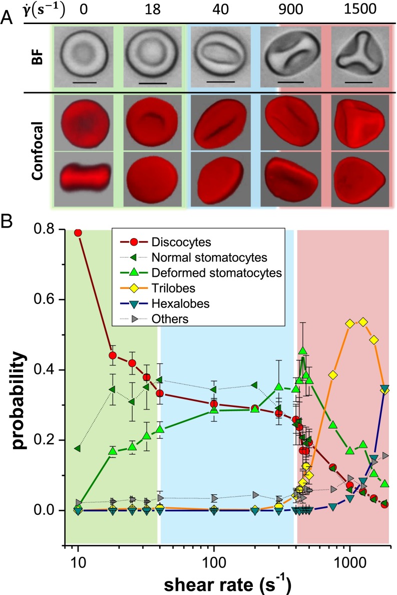
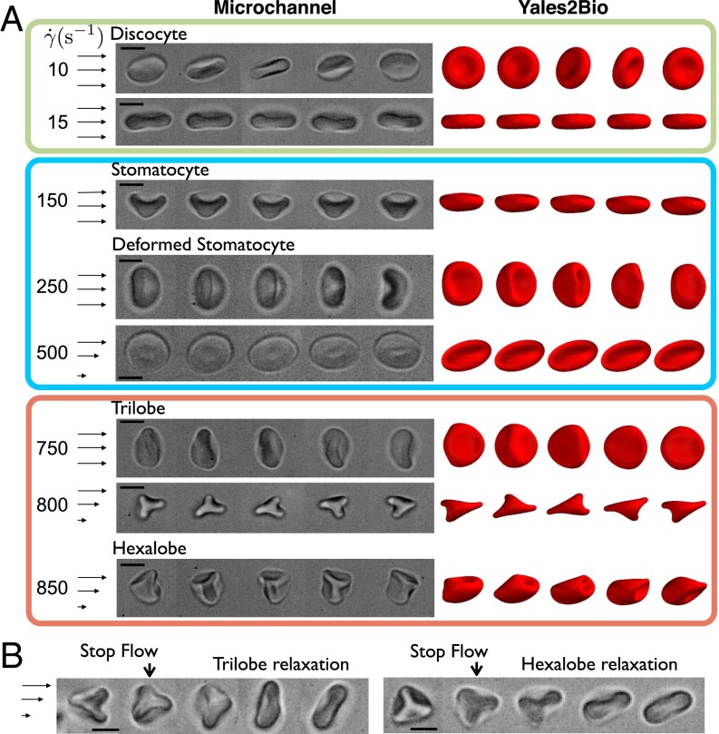
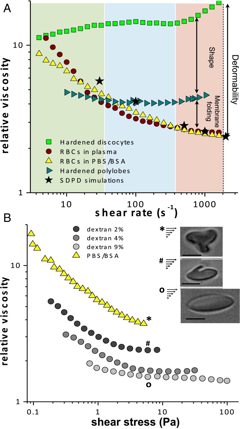
Blood viscosity decreases with shear stress, a property essential for an efficient perfusion of the vascular tree. Shear thinning is intimately related to the dynamics and mutual interactions of RBCs, the major component of blood. Because of the lack of knowledge about the behavior of RBCs under physiological conditions, the link between RBC dynamics and blood rheology remains unsettled. We performed experiments and simulations in microcirculatory flow conditions of viscosity, shear rates, and volume fractions, and our study reveals rich RBC dynamics that govern shear thinning. In contrast to the current paradigm, which assumes that RBCs align steadily around the flow direction while their membranes and cytoplasm circulate, we show that RBCs successively tumble, roll, deform into rolling stomatocytes, and, finally, adopt highly deformed polylobed shapes for increasing shear stresses, even for semidilute volume fractions of the microcirculation. Our results suggest that any pathological change in plasma composition, RBC cytosol viscosity, or membrane mechanical properties will affect the onset of these morphological transitions and should play a central role in pathological blood rheology and flow behavior.

Keywords: blood rheology; blood simulation; cell dynamics; red blood cell.


Conflict of interest statement

The authors declare no conflict of interest.

Figures

Fig. 1.
Fig. 1.
Investigation of RBC shapes at different shear rates in a cone-and-plate rheometer. (A) Observation of hardened cells by optical (black and white) and confocal (red) microscopy: With increasing γ˙ the formation of highly deformed stomatocytes and then polylobed cells (trilobe and hexalobe) is detected. (Scale bars, 5 μm.) (B) Shape distribution of RBC populations in samples hardened at different shear rates: the three regions in color highlight different regimes of decrease of discocyte populations. The error bars represent triplicate measurements realized in the two rapidly varying regions. They illustrate the typical variance of the measurements.
Fig. 2.
Fig. 2.
Microfluidic observations of RBC dynamics in shear flow. (A) Sequence of RBC deformationsat various γ˙: time lapse = 20 ms (γ˙=10 s−1), 6 ms (γ˙=15 s−1), 3.25 ms (γ˙=150,250,and 500 s−1), 0.6 ms (γ˙=750 s−1), 1.75 ms (γ˙=800 s−1), and0.6 ms (γ˙=850 s−1). The right side of the figure shows analogous time sequences of RBCs obtained withYALES2BIO simulations. Time lapses are given in 1/γ˙ units from top to bottom: 8, 7, 6, 6, and 7 forthe four last cases. (B) Stop-flow sequences of (Left) a trilobe with a relaxation time of1 s and intermediate images separated by 0.23 and 0.56 s and (Right) a hexalobe with relaxation of 1.2 s and successive images with a time interval of 0.32 and 0.71 s. (Scale bars, 5 μm.)
Fig. 3.
Fig. 3.
Shape distributions of HRBCs at 900 s−1 in suspensions with different Ht. For clarity, discocyte and stomatocyte number densities have been omitted because they are negligible. (Inset) Black frames on the right, a top view and a cross-section of a creased discocyte acquired by confocal microscopy as well as an image of a multilobe in BF. Physiological Hts are indicated by the light yellow region. (Scale bar, 5 μm.)
Fig. S1.
Fig. S1.
Distributions of RBC extension for different Ht values measured in SDPD simulations. Extension is defined as the maximum instantaneous cell length l in the flow direction, as depicted in the first inset. Distributions include data from multiple RBCs in a suspension and are averaged over many time instances. Insets illustrate typical shapes for different values of l. The two extension regions marked by light yellow and light blue colors schematically depict l ranges where polylobed shapes and creased discocytes are observed.
Fig. 4.
Fig. 4.
Rheology of dense suspensions of RBCs (Ht=45%). (A) Relative viscosity of the suspensions of deformable RBCs in plasma (red) and in PBS/BSA (yellow) as a function of γ˙ in comparison with the suspensions of washed cells hardened at rest (green) and at γ˙=1,500 s−1 (blue). Suspensions of deformable RBCs show a typical shear thinning for increasing γ˙, whereas hardened samples have a nearly Newtonian behavior. SDPD simulation data (black stars) are also shown for deformable cells and agree well with the experimental results for RBCs suspended in plasma or in PBS/BSA. (B) Rheology of washed blood in comparison with the suspensions of RBCs in solutions with different dextran concentrations (temperature = 25 °C). (Inset) The effect of viscosity ratio λ is highlighted by the illustration of single cells flowing in both PBS/BSA and dextran solutions in microfluidics at a comparable shear stress τ. (Scale bars, 5 μm.)

References

    1. Wells RE, Merrill EW. Shear rate dependence of the viscosity of whole blood and plasma. Science. 1961;133:763–764. - PubMed
    1. Dintenfass L. Internal viscosity of the red cell and a blood viscosity equation. Nature. 1968;219:956–958. - PubMed
    1. Chien S. Shear dependence of effective cell volume as a determinant of blood viscosity. Science. 1970;168:977–979. - PubMed
    1. Brust M, et al. Rheology of human blood plasma: Viscoelastic versus Newtonian behavior. Phys Rev Lett. 2013;110:078305. - PubMed
    1. Merrill EW, Gilliland ER, Lee TS, Salzman EW. Blood rheology: Effect of fibrinogen deduced by addition. Circ Res. 1966;18:437–446. - PubMed

Publication types

LinkOut - more resources