. 2016;30(12):1399-1413.
doi: 10.1080/02699052.2016.1219058.

Progression of tau pathology within cholinergic nucleus basalis neurons in chronic traumatic encephalopathy: A chronic effects of neurotrauma consortium study

Affiliations

Progression of tau pathology within cholinergic nucleus basalis neurons in chronic traumatic encephalopathy: A chronic effects of neurotrauma consortium study

Elliott J Mufson et al. Brain Inj. 2016.

Abstract

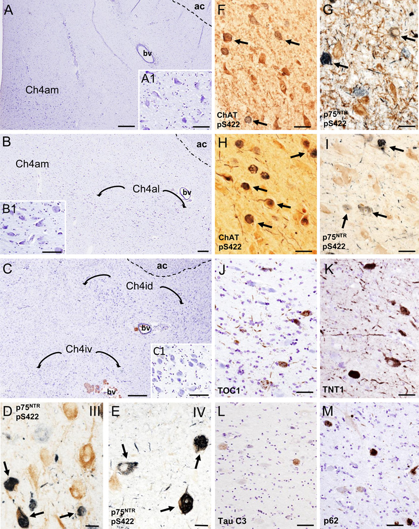
Objective: To test the hypothesis that the nucleus basalis of Meynert (nbM), a cholinergic basal forebrain (CBF) cortical projection system, develops neurofibrillary tangles (NFTs) during the progressive pathological stages of chronic traumatic encephalopathy (CTE) in the brain of athletes.

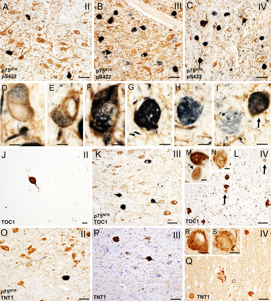
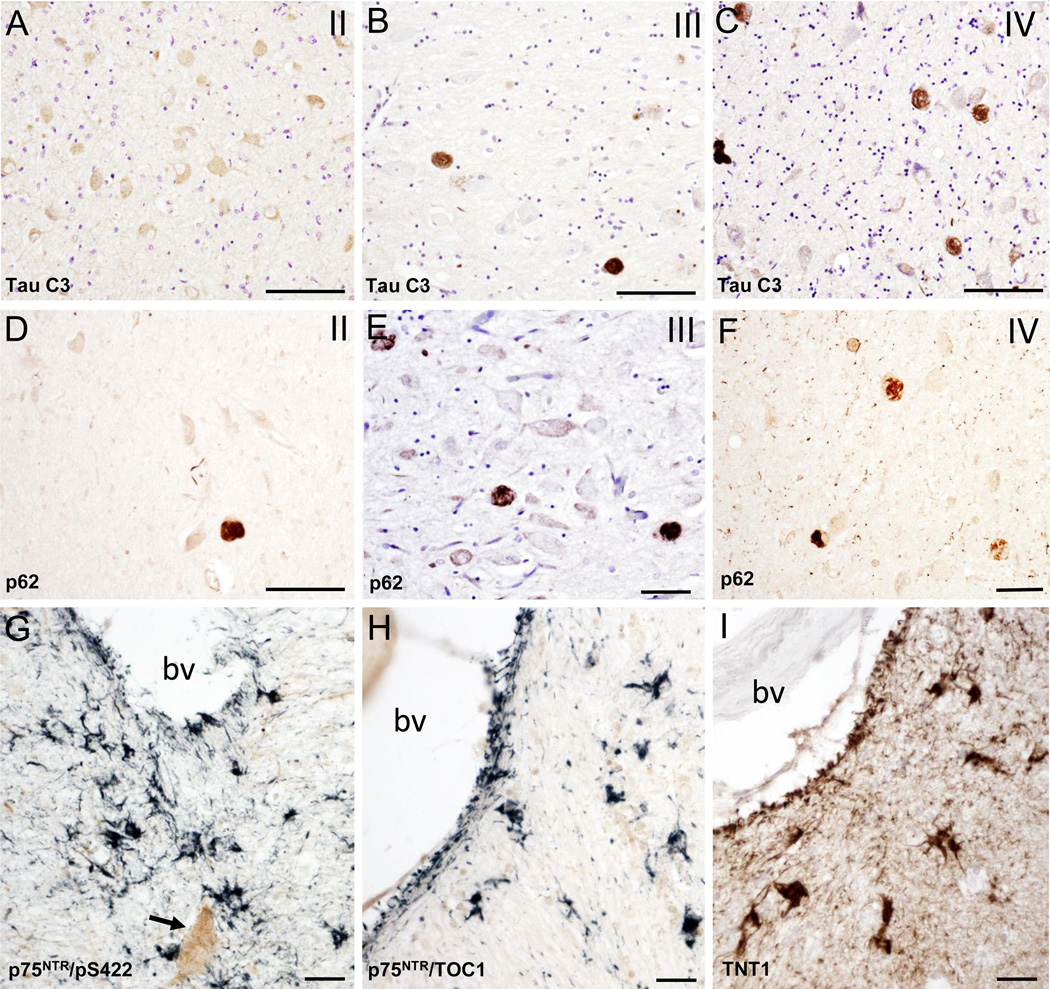
Method: To characterize NFT pathology, tau-antibodies marking early, intermediate and late stages of NFT development in CBF tissue obtained at autopsy from eighteen former athletes and veterans with a history of repetitive mild traumatic brain injury (TBI) were used.

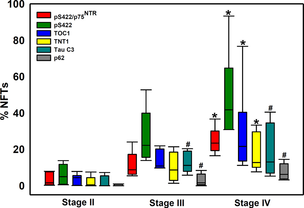
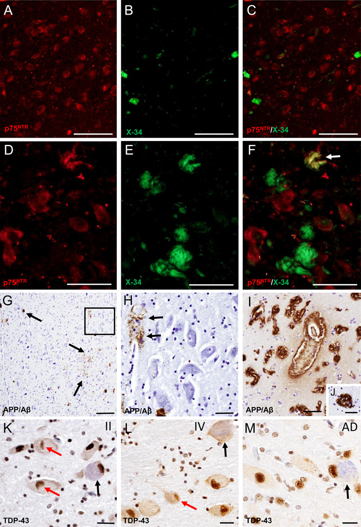
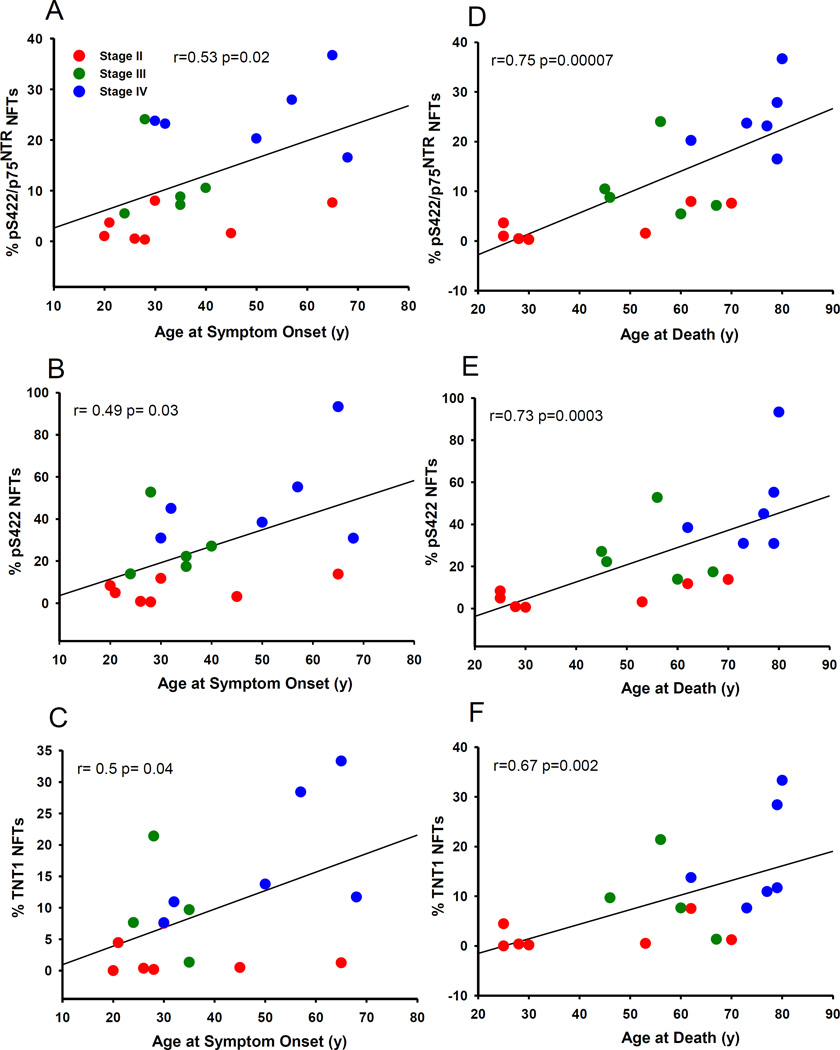
Results: Analysis revealed that cholinergic nbM neurons develop intracellular tau-immunoreactive changes progressively across the pathological stages of CTE. In particular, there was an increase in pre-tangle (phosphorylated pS422) and oligomeric (TOC1 and TNT1) forms of tau in stage IV compared to stage II CTE cases. The nbM neurons also displayed pathologic TDP-43 inclusions and diffuse extracellular and vascular amyloid-β (Aβ) deposits in CTE. A higher percentage of pS422/p75NTR, pS422 and TNT1 labelled neurons were significantly correlated with age at symptom onset, interval between symptom onset and death and age at death.

Conclusion: The development of NFTs within the cholinergic nbM neurons could contribute to an axonal disconnection in CTE. Further studies are needed to determine the mechanism driving NFT formation in the nbM neurons and its relation to chronic cognitive dysfunction in CTE.

Keywords: Cognition; head injury; memory; mild brain injury; traumatic brain injury.


Figures

Figure 1
Figure 1
Photomicrographs of cresyl violet stained sections delineating the cholinergic subfields (Ch4) examined within the nucleus basalis of Meynert (nbM) from rostral to caudal: anteromedial (Ch4am) (A), anterolateral (Ch4al) (B) and dorsal (Ch4id) and ventral (Ch4iv) intermediate (C); A1, B1 and C1insets showing higher magnification images of Ch4am, Ch4al and Ch4id neurons from panels A, B and Crespectively. D and E. Dual immunolabeled p75NTR/pS422 neurons (dark blue; arrows) and single labeled p75NTR (brown) neurons in the vertical limb of the diagonal band of Broca (VDB; Ch2) in a CTE stage III (D) and IV (E) case, respectively. F and G. Photomicrographs of ChAT (F) (brown) and p75NTR-positive (brown) (G) neurons containing pS422-ir inclusions (dark/light blue; arrows) within the nbM anteromedial subfield in CTE stage IV. H and I. Images of the anteromedial subfield of the nbM showing dual immunolabeled ChAT (brown)/pS422 positive neurons (dark blue; arrows) and single immunolabeled ChAT neurons (brown) (H) and double immunolabeled p75NTR /pS422 positive neurons (dark and light blue; arrows) and single-labeled p75NTR neurons (brown) (I) from an AD case, respectively. J-M. Photomicrographs of TOC1 (J), TNT1 (K), Tau C3 (L) and p62 (M) positive neurons and neuropil threads in the anterior subfields of the nbM of an AD case. Note the extent of oligomer TOC1 and TNT1 positive threads compared to absence of neuropil thread Tau C3 and p62 immunostaining. Tissues were counterstained with cresyl violet in panels J to M. Abbreviations: ac, anterior commissure; bv, blood vessel. Scale bars: 500 µm (A), 250 µm (B), 200 µm (C), 100 µm (B1, C1), 80 µm (A1), 75 µm (J, L, M), 60 µm (F), 50 µm (H, I, K), 40 µm (G) and 30 µm (D, E).
Figure 2
Figure 2
A–C. Photomicrographs of tissue dual immunostained for p75NTR (brown) and pS422 (dark blue/black) in the anterior subfields of the nbM showing the presence p75NTR containing pS422 inclusions (dark blue) and neuropil threads (dark blue) in CTE stages II (A), III (B) and IV (C). Note that many more double p75NTR/pS422 positive neurons and neuropil threads (dark blue) were seen in stage III and IV compared to stage II. D–G. High power images of double immunolabeled p75NTR/pS422 neurons in the anteromedial nbM showing intraneuronal cytoplasmic pS422 imunoreactivity (black) containing granular (D and E), filamentous (F) and skein of yarn-like (G) phenotypes in a stage IV CTE case. H and I. Twisted filamentous p75NTR positive inclusion (blue) resembling a skein of yarn in the nbM neuropil. Arrows indicates a double immunolabeled p75NTR/pS422 neuron in I. J–L. Photomicrographs of single TOC1 (brown) (J and L) and dual immunolabeled p75NTR/TOC1 (dark blue/black) neurons and neuropil threads in anterior subfields of the nbM in a stage II (J), III (K) and IV (L) CTE cases. Note the increase in the extent of TOC positive neuropil threads in stage IV. M and N. Insets showing higher magnification images of granular (small arrow), compact spherical (M) and twisted-filamentous (N) TOC1 immunoreactive profiles (brown) in the anterormedial subfield of the nbM neurons from panel L (arrows). O–Q. Photomicrographs of double immunolabeled TNT1/p75NTR (dark blue) (O) and single reactive TNT1 (brown) (P and Q) positive neurons and neuropil threads in the anterior subfields of the nbM from a stage II (O), III (P) and IV (Q) CTE cases. R and S. Insets showing details of the granular and filamentous TOC1 immunoreactivity in anteromedial subfield of the nbM neurons from a stage IV CTE case. Tissue was counterstained with cresyl violet in panel P. Scale bars: 75 µm (A–C), 65 µm (L, O, Q), 50 µm (K, P), 25 µm (J), 15 µm (M, R, S), 12 µm (G), 10 µm (D, E, H) and 8 µm (F, I, N).
Figure 3
Figure 3
Photomicrographs of Tau C3 (A–C) and p62 (D–E) immunolabeling showing many more globose Tau C3 and p62-postive neurons in the anterior subfields of the nbM in a CTE stage IV than stage II. Note the lack of nbM Tau C3 positive neurons in stage II. Photomicrographs of the double immunolabeling for pS422/p75NTR (G), TOC1/p75NTR (H) and single immunolabeling for TNT1 (I) showing pS422 (blue), TOC1 (blue) and TNT1 (brown) immunostained astrocytes near to a blood vessel in anteromedial subfield of the nbM in a CTE stage IV case. Arrow indicates a p75NTR positive cholinergic neuron. Abbreviation: bv, blood vessel. Tissues in panels A, B and E were counterstained with cresyl violet. Scale bars: 90 µm (A–D), 70 µm (E, F), 40 µm (G) and 25 µm (H, I).
Figure 4
Figure 4
Box plot showing the percent of double pS422/p75NTR as well as single pS422, TOC1, TNT1, Tau C3 and p62 immunopositive neurofibrillary tangle (NFTs) relative to the total number of p75 NTR positive neurons in nbM in stages II, III and IV. Percentage of pS422/p75NTR, pS422, TOC1, TNT1positive NFTs were significantly increased in stage IV compared to stage II (*, p<0.01), while the percentage of Tau C3 and p62 NFTs were significantly higher in stage IV and III compared to stage II (#, p<0.01).
Figure 5
Figure 5
A–F. Fluorescence images of single p75NTR (red) (A and D) and X-34 (green) (B and E) staining and merged-images (C and F) showing limited of X-34 positive neurons (B) and the absence of double p75NTR/X-34 positive neurons (C) in the nbM in a CTE stage II case, while many more fibrillary X-34 positive neurons (E and F) were seen in the anterior subfields of the nbM in a CTE stage IV case. Arrow in F indicates a double immunolabeled p75NTR/X-34 neuron (yellow). G. Low magnification image of scattered APP/Aβ (6E10) labeled deposits (brown; arrows) in the nbM anteromedial subfield of a CTE stage IV case. H. High power image of APP/Aβ-positive blood vessel (arrows) from the area boxed in panel G; note the lack of intraneuronal APP/Aβ immunostaning in the large hyperchromic nbM neurons (blue). I–M. Images showing APP/Aβ positive deposits in blood vessels (bv) in the insular cortex as well as APP/Aβ reactive cored neuritic plaques in the temporal cortex (J) from a stage IV CTE case. N. K–M. Images showing nuclear TDP-43 staining (brown) in the nbM neurons from a CTE stage II (K), IV (L) and an AD case (M). Note the presence of lenticular shaped TPD-43-ir inclusions (red arrows) within nbM neurons in a CTE stage II (K) and IV (L), but not in the AD case (M). Black arrows indicate NFT-containing neurons (light blue) in CTE and AD cases. Sections in panels K–M were counterstained with hematoxylin. Scale bars: 200 µm (G), 100 µm (A–C), 50 µm (D–H, J), 40 µm (I), 25 µm (K–M)
Figure 6
Figure 6
Percent of pS422/p75NTR (A and D), pS422 (B and E) and TNT1 (C and F) positive NFTs in the nbM correlated positively with the age of symptom onset (A–C) and age at death (D–F) across CTE stages. Stage II, red circles; Stage III, green circles; Stage IV, blue triangles.

References

    1. Cifu DX, Taylor BC, Carne WF, Bidelspach D, Sayer NA, Scholten J, Campbell EH. Traumatic brain injury, posttraumatic stress disorder, and pain diagnoses in OIF/OEF/OND Veterans. J Rehabil Res Dev. 2013;50:1169–1176. - PubMed
    1. David X Cifu D-AR, Williams RL, Carne W, West SL, McDougal M, Dixon K. Chronic Effects of Neurotrauma Consortium: An Overview at Year 1. Fed Pract. 2015;32:44–48. - PMC - PubMed
    1. Shively S, Scher AI, Perl DP, Diaz-Arrastia R. Dementia resulting from traumatic brain injury: what is the pathology? Arch Neurol. 2012;69:1245–1251. - PMC - PubMed
    1. Shin SS, Dixon CE. Alterations in Cholinergic Pathways and Therapeutic Strategies Targeting Cholinergic System after Traumatic Brain Injury. J Neurotrauma. 2015;32:1429–1440. - PMC - PubMed
    1. Omalu B, Hammers JL, Bailes J, Hamilton RL, Kamboh MI, Webster G, Fitzsimmons RP. Chronic traumatic encephalopathy in an Iraqi war veteran with posttraumatic stress disorder who committed suicide. Neurosurg Focus. 2011;31:E3. - PubMed

MeSH terms

LinkOut - more resources