. 2016 Nov 1;72(Pt 11):820-830.
doi: 10.1107/S2053230X16016149. Epub 2016 Oct 24.

Natural and non-natural amino-acid side-chain substitutions: affinity and diffraction studies of meditope-Fab complexes

Affiliations

Natural and non-natural amino-acid side-chain substitutions: affinity and diffraction studies of meditope-Fab complexes

Krzysztof P Bzymek et al. Acta Crystallogr F Struct Biol Commun. .

Abstract

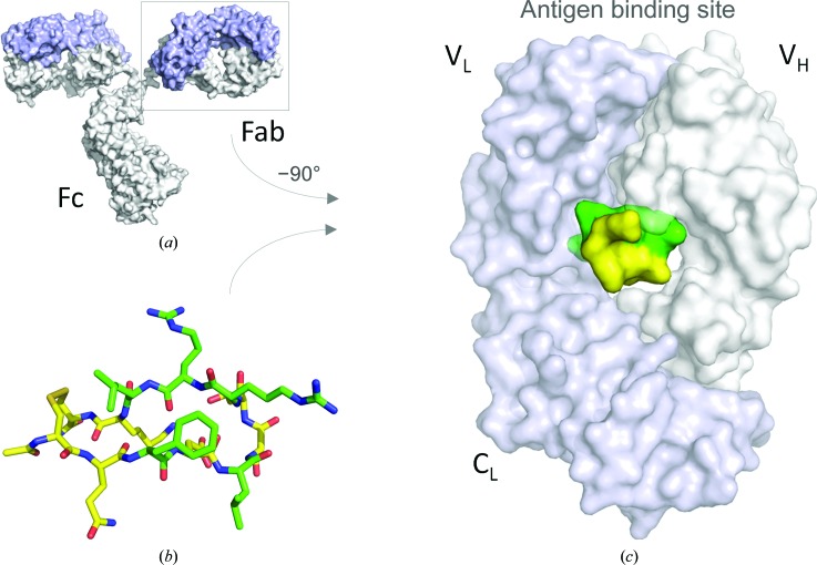
Herein, multiple crystal structures of meditope peptide derivatives incorporating natural and unnatural amino acids bound to the cetuximab Fab domain are presented. The affinity of each derivative was determined by surface plasmon resonance and correlated to the atomic structure. Overall, it was observed that the hydrophobic residues in the meditope peptide, Phe3, Leu5 and Leu10, could accommodate a number of moderate substitutions, but these invariably reduced the overall affinity and half-life of the interaction. In one case, the substitution of Phe3 by histidine led to a change in the rotamer conformation, in which the imidazole ring flipped to a solvent-exposed position. Based on this observation, Phe3 was substituted by diphenylalanine and it was found that the phenyl rings in this variant mimic the superposition of the Phe3 and His3 structures, producing a moderate increase, of 1.4-fold, in the half-life of the complex. In addition, it was observed that substitution of Leu5 by tyrosine and glutamate strongly reduced the affinity, whereas the substitution of Leu5 by diphenylalanine moderately reduced the half-life (by approximately fivefold). Finally, it was observed that substitution of Arg8 and Arg9 by citrulline dramatically reduced the overall affinity, presumably owing to lost electrostatic interactions. Taken together, these studies provide insight into the meditope-cetuximab interaction at the atomic level.

Keywords: X-ray crystallography; cetuximab; meditope; monoclonal antibody; surface plasmon resonance; unnatural amino acids.


Figures

Figure 1
Figure 1
Meditope binding site. (a) Model of cetuximab IgG based on PDB entry 1igt (Harris et al., 1997 ▸). The light chain is shown in light blue and the heavy chain in light gray. (b, c) The cavity in the Fab arm can accommodate the meditope peptide (PDB entry 4gw1; Donaldson et al., 2013 ▸). The residues that are under investigation in this report are highlighted in green.
Figure 2
Figure 2
Tyrosine at position 3 affects Arg8 (stereoviews). (a) Superposition of cQYN (pink C atoms) and F3Y, where the alanine at position 9 (A9) is substituted by Arg (R9) (light blue C atoms), superimposed on the cQFD meditope (green C atoms). The presence of a hydroxyl from Tyr3 (Y3) sterically occludes the Arg8 side chain, resulting in the loss of a hydrogen bond to the backbone of Gln111 in the heavy chain. The hydroxyl group of Y3, however, leads to the coordination of a water molecule. (b) Superposition of the Fab (cQFD in black and cQYN in pink) shows that the hydroxyl substitution leads to a slight reorientation of the meditope with respect to the Fab.
Figure 3
Figure 3
Substitutions of the phenylalanine at position 3 with brominated phenylalanine analogues, shown in stereo and superimposed on the cQFD meditope (green C atoms). (a) Viewed from the top, tyrosine (purple C atoms) at position 3 blocks the extension of the side chain of Arg8 (R8). (b) Substitution with 2-bromophenylalanine (2-BrF; magenta C atoms) does not affect the positioning of R8; however, there are multiple conformation of the 2-BrF side chain. (c) Substitution with 3-bromophenylalanine (3-BrF; yellow C atoms) affects R8; however, it also produces a conformational change in Leu5 (L5). (d) Substitution with 4-bromophenylalanine (4-BrF; blue C atoms) slightly perturbs R8. (e) Side view with each variant superimposed on the cQFD meditope.
Figure 4
Figure 4
Substitutions of the phenylalanine at position 3, shown in stereo and superimposed on the cQFD meditope (green C atoms). (a) Substitution of phenylalanine with glutamine led to multiple side-chain rotamers (hot pink C atoms). (b) Substitution of phenylalanine with histidine led to a single conformation exposed to the solvent (cyan C atoms). (c) Based on these observations, we substituted phenylalanine with diphenylalanine (orange C atoms). One phenyl group of the diphenylalanine substitution superposed with the phenyl ring of the cQFD meditope. The other phenyl group superposed well with the imidazole ring of the histidine meditope variant.
Figure 5
Figure 5
Substitutions of leucine at position 5, shown in stereo and superimposed on the cQFD meditope (green C atoms). (a) The leucine side chain resides in a hydrophobic pocket defined by Thr90 (T90), Ile92 (I92) and Leu114 (L114) of the Fab heavy chain and Pro40 (P40) of the Fab light chain. (b) Substitution of leucine with tyrosine in the meditope positions the hydroxyl group near the side chain of Glu154 (E154) and the hydroxyl group of Tyr182 (Y182). (c) The replacement of leucine with glutamine at position 5 was intended to create a hydrogen bond to the hydroxyl group of Y182 in the heavy chain or the hydroxyl of T90 in the light chain. However, the side chain points away from the Fab. (d) Substitution with glutamic acid resulted in positioning of the carboxylic acid in the hydrophobic pocket. The high B factors of the carboxylate suggest that the positioning of the side chain is adventitious. (e) Substitution at position 5 with diphenylalanine places one phenyl group at the same position as the leucine side chain. The other phenyl group extends further into the meditope cavity that is lined with the hydrophobic residues.
Figure 6
Figure 6
Citrulline substitutions, shown in stereo and superimposed on the cQFD meditope. The substitution of arginine with citrulline at either (a) position 8 or (b) position 9 gave structures that were indistinguishable from that of the original meditope (c).
Figure 7
Figure 7
Substitutions of leucine at position 10, shown in stereo and superimposed on the cQFD meditope. Leu10 packs against a shallow hydrophobic pocket. Glutamine was substituted for Leu10 in an effort to form a hydrogen bond to the hydroxyl group of Tyr87 adjacent to the hydrophobic pocket. The cyan spheres represent two water molecules present in the apo structure (PDB entry 1yy8).
Figure 8
Figure 8
Half-lives of meditope variant–cetuximab interactions. While the on-rate of a bimolecular interaction is dependent on concentration, the off-rate is not. The half-life is related to the off-rate through t = ln(2)/k d. The dashed line represents the lower limit on the determination of k d (0.5 s−1, corresponding to a 1.4 s half-life.) Note that several of the variants were cyclized through a diglycine linker, of which GQ(2-Br-F)DLSTRRLKG [F3(2-BrF)] and GQ(4-Br-F)DLSTRRLKG [F3(4-BrF)] allowed the determination of kinetic constants.

References

    1. Bhakta, S., Raab, H. & Junutula, J. R. (2013). Methods Mol. Biol. 1045, 189–203. - PubMed
    1. Bzymek, K. P., Ma, Y., Avery, K. A., Horne, D. A. & Williams, J. C. (2016). Acta Cryst. F72, 434–442. - PMC - PubMed
    1. Chari, R. V. J. (2008). Acc. Chem. Res. 41, 98–107. - PubMed
    1. Donaldson, J. M., Zer, C., Avery, K. N., Bzymek, K. P., Horne, D. A. & Williams, J. C. (2013). Proc. Natl Acad. Sci. USA, 110, 17456–17461. - PMC - PubMed
    1. Ducry, L. & Stump, B. (2010). Bioconjug. Chem. 21, 5–13. - PubMed

MeSH terms