

Epitope Identification and Application for Diagnosis of Duck Tembusu Virus Infections in Ducks
- PMID: 27834908
- PMCID: PMC5127020
- DOI: 10.3390/v8110306
Epitope Identification and Application for Diagnosis of Duck Tembusu Virus Infections in Ducks
Abstract
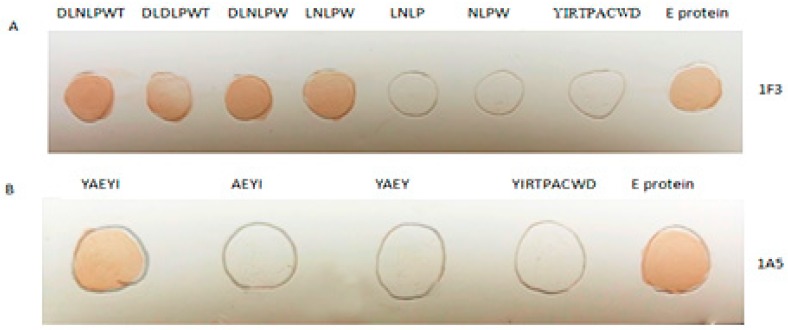
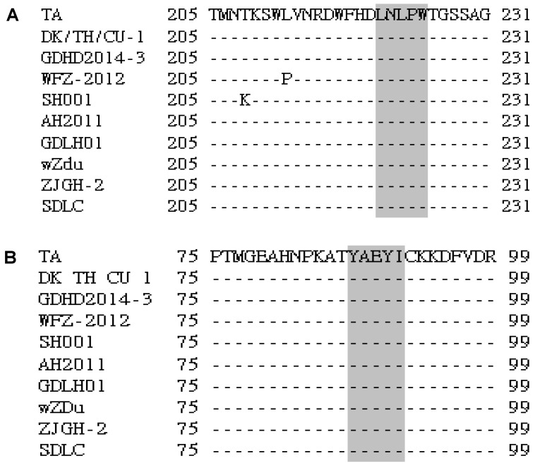
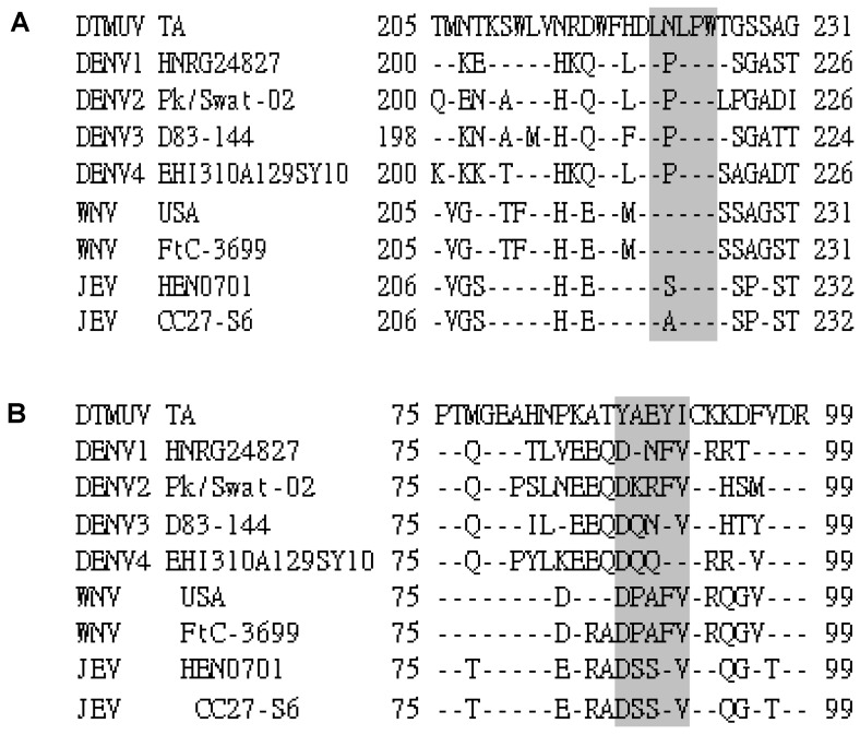
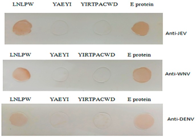
Duck Tembusu virus (DTMUV) causes substantial egg drop disease. DTMUV was first identified in China and rapidly spread to Malaysia and Thailand. The antigenicity of the DTMUV E protein has not yet been characterized. Here, we investigated antigenic sites on the E protein using the non-neutralizing monoclonal antibodies (mAbs) 1F3 and 1A5. Two minimal epitopes were mapped to 221LD/NLPW225 and 87YAEYI91 by using phage display and mutagenesis. DTMUV-positive duck sera reacted with the epitopes, thus indicating the importance of the minimal amino acids of the epitopes for antibody-epitope binding. The performance of the dot blotting assay with the corresponding positive sera indicated that YAEYI was DTMUV type-specific, whereas 221LD/NLPW225 was a cross-reactive epitope for West Nile virus (WNV), dengue virus (DENV), and Japanese encephalitis virus (JEV) and corresponded to conserved and variable amino acid sequences among these strains. The structure model of the E protein revealed that YAEYI and LD/NLPW were located on domain (D) II, which confirmed that DII might contain a type-specific non-neutralizing epitope. The YAEYI epitope-based antigen demonstrated its diagnostic potential by reacting with high specificity to serum samples obtained from DTMUV-infected ducks. Based on these observations, a YAEYI-based serological test could be used for DTMUV surveillance and could differentiate DTMUV infections from JEV or WNV infections. These findings provide new insights into the organization of epitopes on flavivirus E proteins that might be valuable for the development of epitope-based serological diagnostic tests for DTMUV.
Keywords: E protein 3D structure; E protein epitopes; diagnosis; duck Tembusu virus; type specific and cross-reactive epitopes.
Conflict of interest statement
The authors declare no competing financial interests.
Figures


Similar articles
-
Characterization of monoclonal antibodies against duck Tembusu virus E protein: an antigen-capture ELISA for the detection of Tembusu virus infection.Arch Virol. 2015 Mar;160(3):757-64. doi: 10.1007/s00705-014-2312-z. Epub 2015 Jan 15. Arch Virol. 2015. PMID: 25588821
-
Identification of a New Broadly Cross-reactive Epitope within Domain III of the Duck Tembusu Virus E Protein.Sci Rep. 2016 Nov 8;6:36288. doi: 10.1038/srep36288. Sci Rep. 2016. PMID: 27824100 Free PMC article.
-
A Novel Neutralizing Antibody Targeting a Unique Cross-Reactive Epitope on the hi Loop of Domain II of the Envelope Protein Protects Mice against Duck Tembusu Virus.J Immunol. 2020 Apr 1;204(7):1836-1848. doi: 10.4049/jimmunol.1901352. Epub 2020 Mar 4. J Immunol. 2020. PMID: 32132180 Free PMC article.
-
Innate immune responses to duck Tembusu virus infection.Vet Res. 2020 Jul 8;51(1):87. doi: 10.1186/s13567-020-00814-9. Vet Res. 2020. PMID: 32641107 Free PMC article. Review.
-
Duck egg drop syndrome virus: an emerging Tembusu-related flavivirus in China.Sci China Life Sci. 2013 Aug;56(8):701-10. doi: 10.1007/s11427-013-4515-z. Epub 2013 Aug 7. Sci China Life Sci. 2013. PMID: 23917842 Review.
Cited by
-
Oral Vaccination with a DNA Vaccine Encoding Capsid Protein of Duck Tembusu Virus Induces Protection Immunity.Viruses. 2018 Apr 6;10(4):180. doi: 10.3390/v10040180. Viruses. 2018. PMID: 29642401 Free PMC article.
-
Advancements in Research on Duck Tembusu Virus Infections.Viruses. 2024 May 20;16(5):811. doi: 10.3390/v16050811. Viruses. 2024. PMID: 38793692 Free PMC article. Review.
-
Mapping a Type-specific Epitope by Monoclonal Antibody against VP3 Protein of Duck Hepatitis A Type 1 Virus.Sci Rep. 2017 Sep 7;7(1):10820. doi: 10.1038/s41598-017-10909-7. Sci Rep. 2017. PMID: 28883462 Free PMC article.
-
Identification of a Neutralizing Monoclonal Antibody That Recognizes a Unique Epitope on Domain III of the Envelope Protein of Tembusu Virus.Viruses. 2020 Jun 15;12(6):647. doi: 10.3390/v12060647. Viruses. 2020. PMID: 32549221 Free PMC article.
-
Screening and identification of B-cell epitopes within envelope protein of tembusu virus.Virol J. 2018 Sep 17;15(1):142. doi: 10.1186/s12985-018-1052-1. Virol J. 2018. PMID: 30223850 Free PMC article.
References
-
- Lindenbach B.D., Thiel H.J., Rice C.M. Flaviviridae: The viruses and their replication. In: Knipe D.M., Howley P.M., Griffin D.E., Lamb R.A., Martin M.A., Roizman B., Straus S.E., editors. Fields Virology. 5th ed. Lippincott Williams & Wilkins; Philadelphia, PA, USA: 2007. pp. 1101–1152.
Publication types
MeSH terms
Substances
LinkOut - more resources
Full Text Sources
Other Literature Sources
Molecular Biology Databases
Research Materials
