. 2016 Nov 22;7(47):76437-76452.
doi: 10.18632/oncotarget.13261.

Stimulus-dependent dissociation between XB130 and Tks5 scaffold proteins promotes airway epithelial cell migration

Affiliations

Stimulus-dependent dissociation between XB130 and Tks5 scaffold proteins promotes airway epithelial cell migration

Serisha Moodley et al. Oncotarget. .

Abstract

Repair of airway epithelium after injury requires migration of neighboring epithelial cells to injured areas. However, the molecular mechanisms regulating airway epithelial cell migration is not well defined. We have previously shown that XB130, a scaffold protein, is required for airway epithelial repair and regeneration in vivo, and interaction between XB130 and another scaffold protein, Tks5, regulates cell proliferation and survival in human bronchial epithelial cells. The objective of the present study was to determine the role of XB130 and Tks5 interaction in airway epithelial cell migration. Interestingly, we found that XB130 only promotes lateral cell migration, whereas, Tks5 promotes cell migration/invasion via proteolysis of extracellular matrix. Upon stimulation with EGF, PKC activator phorbol 12, 13-dibutyrate or a nicotinic acetylcholine receptor ligand, XB130 and Tks5 translocated to the cell membrane in a stimulus-dependent manner. The translocation and distribution of XB130 is similar to lamellipodial marker, WAVE2; whereas Tks5 is similar to podosome marker, N-WASP. Over-expression of XB130 or Tks5 alone enhances cell migration, whereas co-expression of both XB130 and Tks5 inhibits cell migration processes and signaling. Furthermore, XB130 interacts with Rac1 whereas Tks5 interacts with Cdc42 to promote Rho GTPase activity. Our results suggest that dissociation between XB130 and Tks5 may facilitate lateral cell migration via XB130/Rac1, and vertical cell migration via Tks5/Cdc42. These molecular mechanisms will help our understanding of airway epithelial repair and regeneration.

Keywords: Pathology Section; SH3PXD2A; actin filament associate protein 1-like 2; lamellipodia; lung repair; podosomes.


Conflict of interest statement

CONFLICTS OF INTEREST

There is no conflict of interest.

Figures
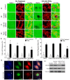
Figure 1
Figure 1. XB130, unlike Tks5, does not play a role in podosome formation and ECM degradation
A.-D. Fluorescent gelatin zymography assay of BEAS-2B cells transfected with control, XB130 or Tks5 siRNA and treated with 500 nM phorbol 12, 13-dibutyrate (PDBu) for 8 h. Cells were stained with rhodamine phalloidin for F-actin (red). Cells were unable to degrade gelatin (green) under basal conditions. PDBu stimulation induced podosome formation (F-actin rich puncta) and gelatin degradation (dark areas). After PDBu treatment, only Tks5 siRNA transfected cells were unable to form podosomes and degrade the gelatin. XB130 siRNA transfected cells behaved like control and control siRNA transfected cells with no effect on the formation or degradation of the gelatin matrix. E. Tks5 downregulation significantly reduces the percentage of cells displaying podosomes, after PDBu treatment, whereas XB130 downregulation did not significantly alter percentage of cells with podosomes. F. Tks5 downregulation significantly reduces the percentage of cells that are able to degrade gelatin, after PDBu treatment. In contrast to Tks5, XB130 downregulation did not alter the percentage of cells able to degrade gelatin. Data is summarized from three independent experiments and presented as mean ± SD. *p < 0.01 compared with controls (non-transfected BEAS-2B cells and non-targeting siRNA-transfected cells). G. Co-immunofluorescence staining of XB130 (green), actin (blue) and Tks5 (red). BEAS2B cells were treated with or without 500 nM PDBu. No treatment control shows normal stress fibers. PDBu treatment shows formation of podosomes (white arrows) as detected by actin and Tks5. XB130 only localizes to the cytoplasm and cell periphery. H. Validation of XB130 or Tks5 siRNA transfection using western blot detection of XB130 and Tks5. Tubulin was used as a representative housekeeping protein.
Figure 2
Figure 2. Tks5 is not essential for lamellipodia formation
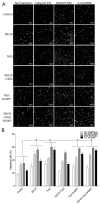
A.-D. BEAS-2B cells were transfected with control (scrambled), XB130 or Tks5 siRNA and subjected to a wound-healing assay over an 8 h time course. Unlike XB130 down regulation, Tks5 down regulation does not inhibit wound closure. E. Percentage of original wound width per hour shows that only XB130 siRNA significantly reduces wound healing at 7 h and 8 h, as compared to control, control siRNA-transfected or Tks5 siRNA-transfected cells. F. High magnification phase contrast microscopy at the leading edge of wounds shows that control and Tks5 downregulated cells form dark ruffled edges (arrow), indicative of lamellipodia, whereas XB130 down-regulated cells appear to lack these structures (asterisk). G. Quantification of cells with lamellipodia at the leading edge shows that XB130 downregulation significantly reduced the percentage of cells displaying lamellipodia, as observed by phase contrast microscopy. Data is summarized from three independent experiments and presented as mean ± SD. * represents p < 0.01 compared with controls (non-transfected BEAS-2B cells and non-targeting siRNA-transfected cells). H. Co-immunofluorescence staining of XB130 (green), actin (blue) and Tks5 (red). BEAS2B cells were treated with or without 50 ng/mL EGF or 0.1 uM NNK. No treatment control shows normal stress fibers. EGF stimulation shows formation of lamellipodia as detected by actin bands at the cell periphery and XB130 staining. NNK stimulation shows the formation of lamellipodia and podosomes (white arrows) as detected by actin and Tks5. XB130 only translocates to lamellipodia after stimulation.
Figure 3
Figure 3. Stimulus-dependent translocation of endogenous XB130 and Tks5 to the cell membrane indicates distinct signaling roles
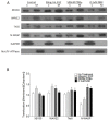
A. Immunoblots of cytoplasm (C) and membrane (M) fractionated BEAS-2B cell lysates. Cells were treated with or without 50 ng/mL EGF, 500 nM PDBu or 0.1 μM NNK. XB130 and WAVE2 expression and translocation from the cytoplasm to the cell membrane are more dependent on EGF stimulation, whereas, Tks5 and N-WASP expression and translocation are more dependent on PDBu and NNK stimulation. B. Ratio of normalized membrane expression to normalized cytoplasm expression. Expression of Na+/K+ ATPase was used to normalize membrane fractions and expression of GAPDH was used to normalize cytoplasmic fractions. Data is summarized from three independent experiments and presented as mean ± SD. * represents p < 0.01 compared to the corresponding no treatment group.
Figure 4
Figure 4. XB130 colocalizes robustly with WAVE2 at lamellipodia but not at podosomes with N-WASP, after stimulation
A.-B. Co-immunofluorescence staining of XB130 (green), actin (blue) and either WAVE2 (A) or N-WASP (B) (red). BEAS2B cells were treated with or without 50 ng/mL EGF, 500 nM PDBu or 0.1 μM NNK. No treatment control shows normal stress fibers. Stimulation with EGF, PDBu and NNK produces actin-rich ruffled areas at the cell membrane, which are indicative of lamellipodia via WAVE2 staining (A). These areas are also enriched with XB130 (A and B). PDBu and NNK induce formation of podosomes (white arrows) which are enriched by N-WASP but not XB130 (B). D. Mander's overlap co-efficient (MOC) of the cell periphery displays the relative colocalization of XB130 with WAVE2, Tks5 or N-WASP, where 0 represents no colocalization and 1 represents perfect colocalization. XB130 colocalizes robustly with WAVE2 at the lamellipodia and to a lesser extent with Tks5 and N-WASP, indicating it translocates to and is involved in lamellipodia formation. Data is summarized from 10 different cells per group from 3 different experiments and presented as mean ± SD. * represents p < 0.01 for XB130/N-WASP and XB130/Tks5 MOCs compared to XB130/WAVE2 MOCs.
Figure 5
Figure 5. Co-expression of XB130 and Tks5 inhibit the enhanced cell migration observed in cells overexpressing XB130 or Tks5 alone
Double chamber transwell cell migration assay using cells transfected with GFP/mCherry-vectors (Control), GFP-XB130/mCherry, GFP/mCherry-Tks5, GFP-XB130/mCherry-Tks5, GFP/mCherry/Myc-Tks5 SH3#5* or GFP-XB130/mCherry/Myc-Tks5 SH3#5*. Cells were stimulated with 50 ng/mL EGF, 500 nM PDBu or 0.1 μM NNK for 1 h. FBS was added to the lower chamber as a chemoattractant. A. Images of cells that migrated past the transwell membrane. Scale bars represent 500 μm. B. Percentage of migrated cells past the transwell membrane. Over-expression of XB130 or Tks5 alone or Tks5 SH3#5* with or without XB130 significantly increases cell migration, whereas, co-expression of both XB130 and Tks5 has only a moderate increase in cell migration as compared to control cells, after stimulation. Data is summarized from three independent experiments and presented as mean ± SD. * represents p < 0.01 of XB130 or Tks5 over-expressing groups versus the control vector-transfected group.
Figure 6
Figure 6. Co-expression of XB130 with Tks5 inhibits the enhanced ECM-dependent cell migration observed in cells over-expressing Tks5 only
Matrigel-coated double chamber transwell cell migration assay using cells transfected with GFP/mCherry-vectors (Control), GFP-XB130/mCherry, GFP/mCherry-Tks5, and GFP-XB130/mCherry-Tks5. Cells were stimulated with 50 ng/mL EGF, 500 nM PDBu or 0.1 μM NNK for 1 h. FBS was added to the lower chamber as a chemoattractant. A. Images of cells cultured on a thin matrigel coating at 0 μm and migration to 200 μm. Scale bars represent 500 μm. B. Percentage of cells migrating to a depth of 200 μm of matrigel. Over-expression of Tks5 alone or Tks5 SH3#5* with or without XB130 significantly increases cell migration into matrigel, whereas, over-expression of XB130 alone does not significantly increase ECM-dependent cell migration, after stimulation, as compared to control cells. Cells co-expressing XB130 with Tks5 show a similar percentage of migrating cells as XB130 over-expression alone. The over-expression of XB130 appears to inhibit Tks5-mediated cell migration, indicating a regulatory effect of XB130 on Tks5. Data is summarized from three independent experiments and presented as mean ± SD. * represents p < 0.01 of the Tks5 over-expressing groups versus vector-transfected control group.
Figure 7
Figure 7. XB130 over-expression enhances Rac1 activation whereas Tks5 over-expression enhances Cdc42 activation
A. BEAS-2B cells were transfected with GFP/mCherry-vectors (transfection control), GFP-XB130/mCherry vector, GFP/mCherry-Tks5, GFP-XB130 /mCherry-Tks5, GFP/Tks5 SH3#5* or GFP-XB130/Tks5 SH3#5*. Cells were stimulated with or without 50 ng/mL EGF, 500 nM PDBu or 0.1 μM NNK for 1 h. Cell lysates were subjected to GST-PAK-PBD pull-down assay and immunoblotted with Rac1-GTP, Cdc42-GTP, XB130 and Tks5. Total Rac1 and total Cdc42 in cell lysates were blotted for comparison, with GAPDH as a loading control. Rac1-GTP was increased by EGF, PDBu or NNK stimulation, especially in cells over-expressing XB130 alone. Cdc42-GTP increased after PDBu or NNK stimulation and was enhanced by Tks5 over-expression. Rac1-GTP or Cdc42-GTP detection was reduced in cell lysates co-expressing XB130 and Tks5, compared with XB130 or Tks5 alone transfected cells, respectively. Tks5 SH3#5* expression with or without XB130 co-expression rescued RAC-GTP but not Cdc42-GTP expression after stimulation, specifically with EGF. XB130 and Tks5 were only detected in GST-PAK-PBD pull-downs of cells stimulated with EGF, PDBu or NNK, whereas co-expression of XB130 and Tks5 reduced their detection in GST-PAK-PBD pulldowns. B. Co-immunoprecipitation of endogenous XB130 or Tks5 and immunoblots of Rac1, Cdc42, PAK1, Tks5 and XB130. XB130 immunoprecipitation effectively pulls down Rac1 under all treatment conditions. By contrast, immunoprecipitation of Tks5 does not pulldown Rac1, weakly pulls down Cdc42 and more effectively pulls down PAK1. Co-immunoprecipitation between XB130 and Tks5 appears to decrease after stimulation with EGF, PDBu or NNK, especially in the Tks5 immunoprecipitation blot. GAPDH is used as a reference to show the initial input of protein concentration used in each immunoprecipitation reaction.
Figure 8
Figure 8. Schematic diagram of the role of XB130 and Tks5 in Rac1 and Cdc42-associated cytoskeletal remodeling for lung epithelial cell migration
Cell migration requires cytoskeleton remodeling mediated by the Arp2/3 complex, which results in the formation of branched, F-actin rich structures (red ball and sticks), such as lamellipodia and podosomes. This diagram shows the A. Top-down view and B. Side-view of a cell displaying lamellipodia and podosome. We demonstrated a novel mechanism for lung epithelial cell migration, in which extracellular factors stimulate a sub-population of XB130 to dissociate from Tks5 and translocate to the cell periphery to promote Rac1-activated signaling of WAVE2-associated lamellipodia formation for cell extension and Tks5 mediation of Cdc42 activity via PAK1 interaction for the promotion of N-WASP-associated podosome assembly and function for ECM-dependent cell migration. Dashed black lines represent translocation of XB130 and Tks5 to the cell membrane.

Similar articles

Cited by

References

    1. Xiao H, Li DX, Liu M. Knowledge translation: airway epithelial cell migration and respiratory diseases. Cell Mol Life Sci. 2012;69:4149–62. doi: 10.1007/s00018-012-1044-z. - DOI - PMC - PubMed
    1. Belperio JA, Lake K, Tazelaar H, Keane MP, Strieter RM, Lynch JP., 3rd Bronchiolitis obliterans syndrome complicating lung or heart-lung transplantation. Semin Respir Crit Care Med. 2003;24:499–530. doi: 10.1055/s-2004-815601. - DOI - PubMed
    1. Pawson T, Scott JD. Signaling through scaffold, anchoring, and adaptor proteins. Science. 1997;278:2075–80. - PubMed
    1. Flynn DC. Adaptor proteins. Oncogene. 2001;20:6270–2. doi: 10.1038/sj.onc.1204769. - DOI - PubMed
    1. Murphy DA, Courtneidge SA. The ‘ins’ and ‘outs’ of podosomes and invadopodia: characteristics, formation and function. Nat Rev Mol Cell Biol. 2011;12:413–26. doi: 10.1038/nrm3141. - DOI - PMC - PubMed

MeSH terms

Substances