

Herpes Simplex Virus Type 1 Shedding in Tears and Nasal and Oral Mucosa of Healthy Adults
- PMID: 27835628
- PMCID: PMC5117635
- DOI: 10.1097/OLQ.0000000000000522
Herpes Simplex Virus Type 1 Shedding in Tears and Nasal and Oral Mucosa of Healthy Adults
Abstract
Background: Herpes simplex virus type 1 (HSV-1) is prevalent worldwide and causes mucocutaneous infections of the oral area. We aimed to define the frequency and anatomic distribution of HSV-1 reactivation in the facial area in persons with a history of oral herpes.
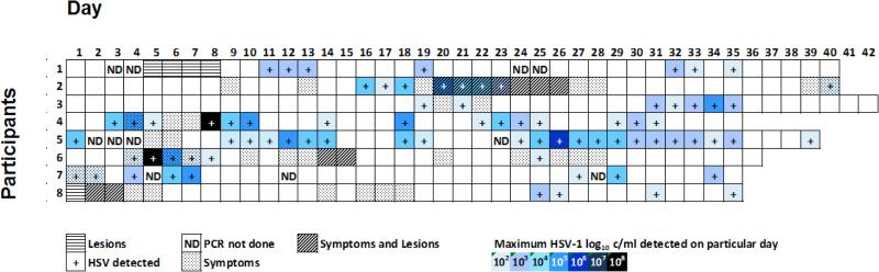
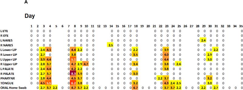
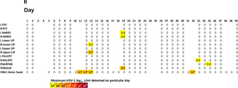
Methods: Eight immunocompetent HSV-1 seropositive adults were evaluated for shedding of HSV-1 from 12 separate orofacial sites (8 from oral mucosa, 2 from nose, and 2 from conjunctiva) 5 days a week and from the oral cavity 7 days a week for approximately 5 consecutive weeks by a HSV DNA PCR assay. Symptoms and lesions were recorded by participants.
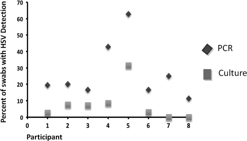
Results: Herpes simplex virus type 1 was detected at least from 1 site on 77 (26.5%) of 291 days. The most frequent site of shedding was the oral mucosa, with widespread shedding throughout the oral cavity. Lesional shedding rate was 36.4% (4 of 11 days with lesions), and the asymptomatic rate was 27.1% (65 of 240 nonlesional days). In individual participants, the median rate of HSV shedding by HSV PCR was 19.7% of days (range, 11%-63%).
Conclusions: Reactivation of HSV-1 on the oral mucosa is common and usually asymptomatic. However, HSV-1 is rarely found in tears and nasal mucosa. Frequent oral shedding of HSV-1 may increase the risk for transmitting the virus to both oral and genital mucosa of sexual partners.
Figures
References
-
- Bradley H, Markowitz LE, Gibson T, McQuillan GM. Seroprevalence of herpes simplex virus types 1 and 2--United States, 1999-2010. The Journal of infectious diseases. 2014;209:325–333. doi:10.1093/infdis/jit458. - PubMed
-
- Tuokko H, Bloigu R, Hukkanen V. Herpes simplex virus type 1 genital herpes in young women: current trend in Northern Finland. Sexually transmitted infections. 2014;90:160. doi:10.1136/sextrans-2013-051453. - PubMed
-
- Coyle PV, et al. Emergence of herpes simplex type 1 as the main cause of recurrent genital ulcerative disease in women in Northern Ireland. Journal of clinical virology : the official publication of the Pan American Society for Clinical Virology. 2003;27:22–29. - PubMed
Publication types
MeSH terms
Grants and funding
LinkOut - more resources
Full Text Sources
Other Literature Sources
Medical
