Review
. 2017 May 1;26(13):700-717.
doi: 10.1089/ars.2016.6942. Epub 2017 Jan 4.

Reactive Oxygen Species/Nitric Oxide Mediated Inter-Organ Communication in Skeletal Muscle Wasting Diseases

Affiliations
Review

Reactive Oxygen Species/Nitric Oxide Mediated Inter-Organ Communication in Skeletal Muscle Wasting Diseases

Lucia M Leitner et al. Antioxid Redox Signal. .

Abstract

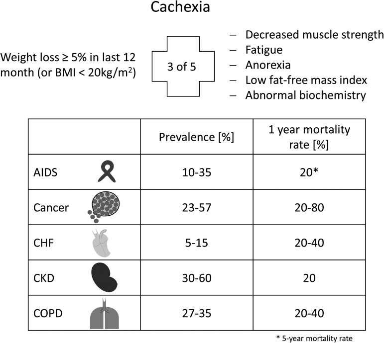
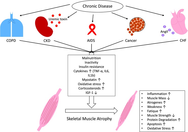
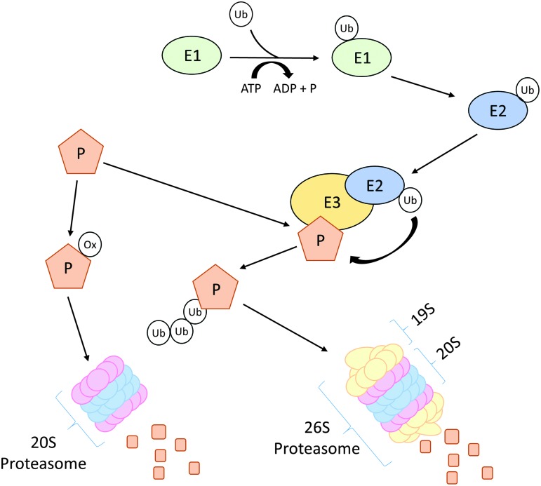
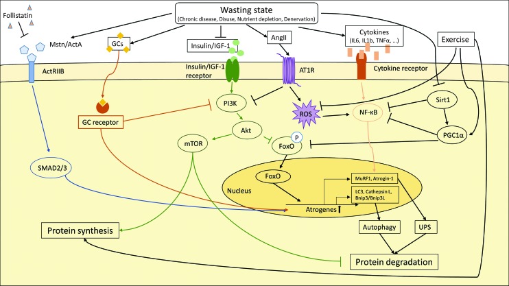
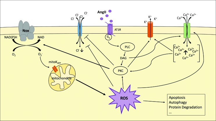
Significance: Cachexia is defined as a complex metabolic syndrome that is associated with underlying illness and a loss of muscle with or without loss of fat mass. This disease is associated with a high incidence with chronic diseases such as heart failure, cancer, chronic obstructive pulmonary disease (COPD), and acquired immunodeficiency syndrome (AIDS), among others. Since there is currently no effective treatment available, cachectic patients have a poor prognosis. Elucidation of the underlying mechanisms is, therefore, an important medical task. Recent Advances: There is accumulating evidence that the diseased organs such as heart, lung, kidney, or cancer tissue secrete soluble factors, including Angiotensin II, myostatin (growth differentiation factor 8 [GDF8]), GDF11, tumor growth factor beta (TGFβ), which act on skeletal muscle. There, they induce a set of genes called atrogenes, which, among others, induce the ubiquitin-proteasome system, leading to protein degradation. Moreover, elevated reactive oxygen species (ROS) levels due to modulation of NADPH oxidases (Nox) and mitochondrial function contribute to disease progression, which is characterized by loss of muscle mass, exercise resistance, and frailty.

Critical issues: Although substantial progress was achieved to elucidate the pathophysiology of cachexia, effectice therapeutic strategies are urgently needed.

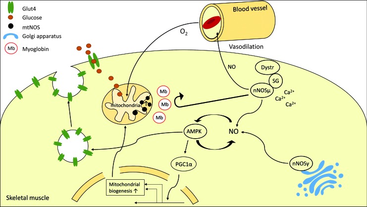
Future directions: With the identification of key components of the aberrant inter-organ communication leading to cachexia, studies in mice and men to inhibit ROS formation, induction of anti-oxidative superoxide dismutases, and upregulation of muscular nitric oxide (NO) formation either by pharmacological tools or by exercise are promising approaches to reduce the extent of skeletal muscle wasting. Antioxid. Redox Signal. 26, 700-717.

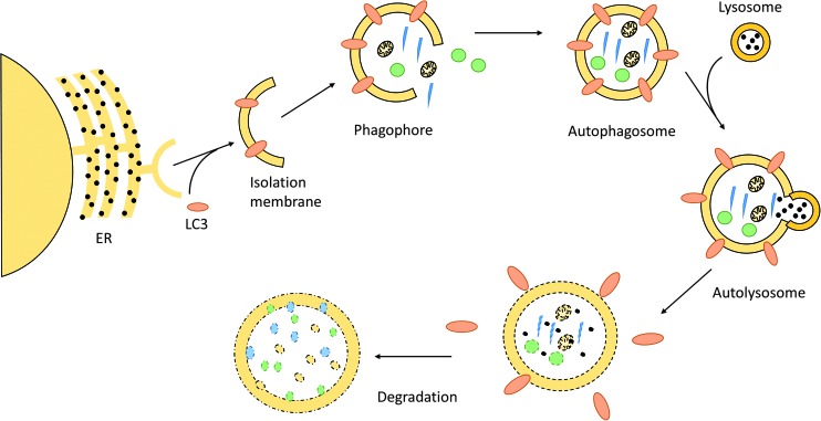
Keywords: angiotensin; autophagy; cachexia; exercise; skeletal muscle; ubiquitin.


Figures

<b>FIG. 1.</b>
FIG. 1.
Definition, prevalence, and mortality rate of cachexia. General parameters to diagnose cachexia and the variability in prevalence and 1-year mortality of cachexia-associated chronic diseases (AIDS*, cancer, CHF, CKD, and COPD). *5-Year mortality rate. CHF, chronic heart failure; CKD, chronic kidney disease; COPD, chronic obstructive pulmonary disease.
<b>FIG. 2.</b>
FIG. 2.
Skeletal muscle wasting induced by chronic diseases. Chronic diseases, such as COPD, CKD, AIDS, cancer, and CHF, share common metabolic changes (malnutrition; inactivity; insulin resistance; increased levels of cytokines [TNFα, IL6, IL1β], myostatin, and corticosteroids; increased oxidative stress; and decreased levels of IGF-1), which lead to skeletal muscle atrophy. Besides these common features, some disease-specific factors further contribute to muscle wasting, for example, uremic toxins in CKD or AngII in CHF. All these metabolic changes induce a catabolic program in skeletal muscles that is indicated by increased inflammation, weakness, fatigue, protein degradation, atrogene expression, apoptosis, and oxidative stress as well as decreased muscle mass and muscle strength. AngII, angiotensin II; IGF-1, insulin-like growth factor 1; IL, interleukin; TNFα, tumor necrosis factor alpha. To see this illustration in color, the reader is referred to the web version of this article at www.liebertpub.com/ars
<b>FIG. 3.</b>
FIG. 3.
Schematic overview of ubiquitin–proteasome pathway. A protein can be marked for degradation by either ubiquitination or oxidation. Activated ubiquitin binds to E1 and is then transferred to E2, the ubiquitin-conjugating enzyme. The loaded E2 delivers the ubiquitin to ubiquitin ligases (E3), which covalently attach ubiquitin to a lysine residues of target proteins (P). Poly-ubiquitinated proteins are then degraded by the 26S proteasome consisting of a 19S regulatory and a 20S core subunit. If a protein gets oxidized (ox), it will be directly degraded via the 20S proteasome. To see this illustration in color, the reader is referred to the web version of this article at www.liebertpub.com/ars
<b>FIG. 4.</b>
FIG. 4.
Schematic overview of autophagy. Autophagy is initiated by Microtubule-associated protein 1A/1B-LC3 conjugation to the nascent phagophore, called isolation membrane—a membrane part derived from the endoplasmic reticulum (ER). While the phagophore expands, it engulfs degradable substrates and finally forms the autophagosome. The autophagosome fuses with a lysosome, which releases its hydrolytic enzymes into the newly formed autolysosome. The content thereby gets degraded and can be recycled by the cell. LC3, microtubule-associated protein 1 light chain 3. To see this illustration in color the reader is referred to the web version of this article at www.liebertpub.com/ars
<b>FIG. 5.</b>
FIG. 5.
Molecular pathways involved in skeletal muscle protein degradation. The induction of atrogenes in catabolic muscle is driven by various molecular pathways. Upregulation of atrogenes leads to skeletal muscle protein degradation via the ubiquitin–proteasome system (UPS) and autophagy. Specific transcription factors such as FoxO proteins, NF-κB and SMAD2/3, as well as GCs activate the transcription of atrogenes. The transcription factors themselves are activated by external stimuli–myostatin (Mstn), activin A (ActA), GCs, insulin, IGF-1, and cytokines. In addition, the anabolic PI3K–AKT–mTOR activity is suppressed, which decreases skeletal muscle protein synthesis and leads to accelerated skeletal muscle protein degradation. Exercise, on the other hand, promotes protein synthesis, blocks ROS production, and enhances PGC1α gene expression. PI3K, phosphoinositide 3; AKT, V-Akt Murine Thymoma Viral Oncogene Homolog 1; mTor, mammalian target of rapamycin; MuRF1, Muscle RING Finger 1; Sirt1, NAD-dependent protein deacetylase sirtuin 1; IL1b/IL6, interleukin 1b/6; TNFα, tumor necrosis factor α; LC3, microtubule-associated protein 1 light chain 3; Bnip3/Bnip3l, BCL2 interacting protein 3/ligand; AT1R, Angiotensin II receptor type 1; ROS, reactive oxygen species; ActRIIb, activin receptor IIb; FoxO, forkhead box protein O; GCs, glucocorticoids; NF-κB, nuclear factor kappa B; PGC1α, peroxisome proliferator-activated receptor gamma coactivator 1-alpha. To see this illustration in color, the reader is referred to the web version of this article at www.liebertpub.com/ars
<b>FIG. 6.</b>
FIG. 6.
Angiotensin II-induced ROS production via NADPH oxidase. AngII binds to its receptor (AT1R) and induces PKC via the Gq-PLC pathway—which is dependent on DAG as well as on increased cytosolic Ca2+ ions. PKC then activates Nox, blocks Cl influx, and induces the K+ export. Nox produces superoxide anions (O2·), which open mitochondrial ATP-dependent potassium channels (mitoKATP). This, subsequently, leads to increased mitochondrial ROS production, which gets released in the cytosol. The increasing amount of ROS further induces PKC and the Ca2+ influx and blocks Cl import. ROS is associated with catabolic mechanism, such as apoptosis, autophagy, and protein degradation. DAG, diacylglycerol; Nox, NADPH oxidase; PKC, proteinkinase C; PLC, phospholipase C. To see this illustration in color, the reader is referred to the web version of this article at www.liebertpub.com/ars
<b>FIG. 7.</b>
FIG. 7.
Native and stained cross-sections of murine hindlimb muscle. Serial 8 μm-thick cryosections of (a) native, (b) SDH-stained, and (c) MyHC-specific-stained skeletal muscles (I: MyHC 1 [red]; IIa: MyHC 2a [green]; IId: MyHC 2d/x [unstained/black]; IIb: MyHC 2b [blue]) revealing the fiber-type distribution as well as their oxidative capacity (SDH stain). Bars = 100 μm. MyHC, myosin heavy chain; SDH, succinate dehydrogenase. To see this illustration in color, the reader is referred to the web version of this article at www.liebertpub.com/ars
<b>FIG. 8.</b>
FIG. 8.
Modulation of skeletal muscle by nitric oxide. nNOSμ associated with the sarcolemma may induce vasodilation, thereby increasing oxygen and nutrient supply by vasodilation. NO enhances AMPK activity and AMPK stimulates NO formation, leading to a feed-forward loop. Activated AMPK, in an NO-dependent manner, promotes translocation of GLUT4 storage vesicles to the sarcolemma, enhancing glucose uptake. Moreover, PGC1α is activated, enhancing mitochondrial biogenesis in skeletal muscle fibers. nNOSγ is associated with the Golgi apparatus. Moreover, a mtNOS has been postulated. The spatial organization of the different NOS compartments may be established by myoglobin, which prevents NO from diffusing between compartments. AMPK, AMP-dependent protein kinase; mtNOS, mitochondrial NOS; nNOS, neuronal NOS; NO, nitric oxide. To see this illustration in color, the reader is referred to the web version of this article at www.liebertpub.com/ars

Similar articles

Cited by

References

    1. MRC/BHF Heart Protection Study of antioxidant vitamin supplementation in 20,536 high-risk individuals: A randomised placebo-controlled trial. Lancet 360: 23–33, 2002 - PubMed
    1. Abrigo J, Rivera JC, Simon F, Cabrera D, and Cabello-Verrugio C. Transforming growth factor type beta (TGF-beta) requires reactive oxygen species to induce skeletal muscle atrophy. Cell Signal 28: 366–376, 2016 - PubMed
    1. Acharyya S, Butchbach ME, Sahenk Z, Wang H, Saji M, Carathers M, Ringel MD, Skipworth RJ, Fearon KC, Hollingsworth MA, Muscarella P, Burghes AH, Rafael-Fortney JA, and Guttridge DC. Dystrophin glycoprotein complex dysfunction: a regulatory link between muscular dystrophy and cancer cachexia. Cancer Cell 8: 421–432, 2005 - PubMed
    1. Adams V, Linke A, Krankel N, Erbs S, Gielen S, Mobius-Winkler S, Gummert JF, Mohr FW, Schuler G, and Hambrecht R. Impact of regular physical activity on the NAD(P)H oxidase and angiotensin receptor system in patients with coronary artery disease. Circulation 111: 555–562, 2005 - PubMed
    1. Adams V, Yu J, Mobius-Winkler S, Linke A, Weigl C, Hilbrich L, Schuler G, and Hambrecht R. Increased inducible nitric oxide synthase in skeletal muscle biopsies from patients with chronic heart failure. Biochem Mol Med 61: 152–160, 1997 - PubMed

Publication types

MeSH terms

LinkOut - more resources