

doi: 10.1186/s12915-016-0322-x.
The evolving world of pseudoenzymes: proteins, prejudice and zombies
Affiliations
- PMID: 27835992
- PMCID: PMC5106787
- DOI: 10.1186/s12915-016-0322-x
Item in Clipboard
The evolving world of pseudoenzymes: proteins, prejudice and zombies
BMC Biol.
.
Abstract
Pseudoenzymes are catalytically deficient variants of enzymes that are represented in all major enzyme families. Their regulatory functions in signalling pathways are shedding new light on the non-catalytic functions of active enzymes, and are suggesting new ways to target cellular signalling mechanisms with drugs.
Figures
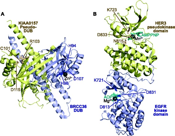
Pseudoenzymes can allosterically regulate a partner protein’s catalytic activity. a The pseudo-deubiquitinase (DUB), KIAA0157 (yellow), which lacks a canonical Zn-binding motif (residues shown as sticks and labelled), binds to the canonical DUB, BRCC36 (blue), to promote its DUB activity. BRCC36 binds Zn2+ (grey sphere) via the labelled residues (shown as blue sticks). The KIAA0157–BRCC36 heterodimeric complex assembles into a higher order “superdimer”, which is an active DUB. PDB accession 5CW3 [11]. b The pseudokinase domain of HER3 (yellow) binds ‘head-to-tail’ and allosterically regulates the activation of the conventional protein tyrosine kinase EGFR (blue). While each domain can accommodate and bind to a nucleotide (cyan) and a divalent cation (grey spheres), HER3 exhibits defective catalytic activity owing to substitution of the catalytic Asp acid residue to an Asn at position 815 (yellow sticks). In contrast, EGFR contains conventional catalytic residues (blue sticks, labelled), which allow it to phosphorylate substrates on tyrosine residues. PDB accession 4RIW [31]. Cartoons were drawn using Pymol
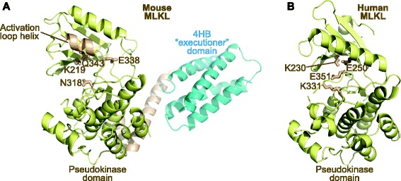
Pseudoenzymes can adopt multiple structural conformations, permitting them to function as molecular switches. a The pseudokinase domain of mouse MLKL was crystallised in an open (equivalent to catalytically inactive) conformation in which the activation loop adopted an unusual helix that buttresses against, and shuns, the αC helix. Counterparts of the catalytic residues in conventional protein kinases (K219, N318, E338), and the K219-interacting Q343 from the activation loop helix, are shown as yellow sticks. PDB accession 4BTF [18]. b The structure of the human MLKL pseudokinase domain crystallised in a distinct closed conformation which resembles that of an active conventional protein kinase, suggesting that MLKL has evolved to function as a catalytically inactive conformational switch. Counterparts of the catalytic residues in conventional protein kinases (K230, K331, E351) are shown as yellow sticks. The canonical αC helix glutamic acid (E250), rather than the activation loop residue observed in the mouse structure, interacts with K230, as is typical of active protein kinase structures. PDB accession 4MWI [14]. Accompanying mutational analyses illustrated that nucleotide binding by MLKL is mediated by different pseudoactive site determinants and that K219 (mouse) and K230 (human) have evolved unexpected functions to permit nucleotide binding, which might drive or inhibit a switch mechanism that controls release of the MLKL four-helix bundle (4HB) domain (shown in a) to induce cell death by necroptosis [14, 18]. Cartoons drawn using Pymol
References
Publication types
MeSH terms
Substances
Grants and funding
LinkOut - more resources
Full Text Sources
Other Literature Sources
