Review
. 2016 Nov-Dec;4(6):1039-1052.
doi: 10.1016/j.jaip.2016.08.005.

Lung Disease in Primary Antibody Deficiencies

Affiliations
Review

Lung Disease in Primary Antibody Deficiencies

Edith Schussler et al. J Allergy Clin Immunol Pract. 2016 Nov-Dec.

Abstract

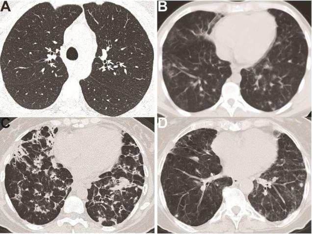
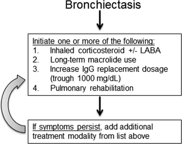
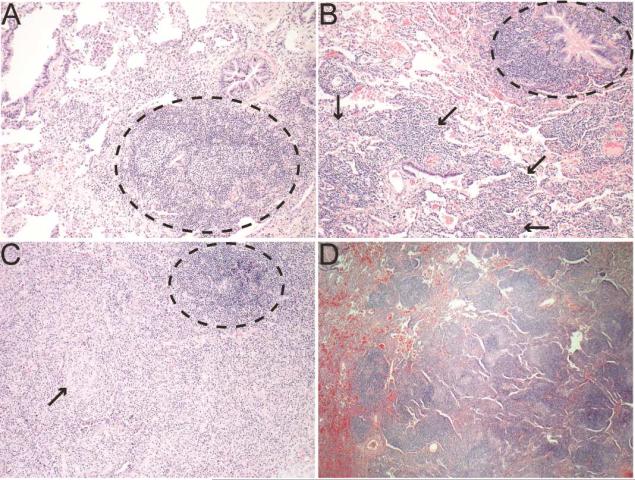
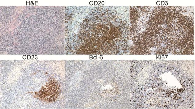
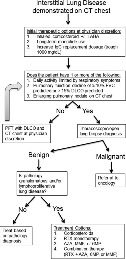
Primary antibody deficiencies (PADs) are the most common form of primary immunodeficiency and predispose to severe and recurrent pulmonary infections, which can result in chronic lung disease including bronchiectasis. Chronic lung disease is among the most common complications of PAD and a significant source of morbidity and mortality for these patients. However, the development of lung disease in PAD may not be solely the result of recurrent bacterial infection or a consequence of bronchiectasis. Recent characterization of monogenic immune dysregulation disorders and more extensive study of common variable immunodeficiency have demonstrated that interstitial lung disease (ILD) in PAD can result from generalized immune dysregulation and frequently occurs in the absence of pneumonia history or bronchiectasis. This distinction between bronchiectasis and ILD has important consequences in the evaluation and management of lung disease in PAD. For example, treatment of ILD in PAD typically uses immunomodulatory approaches in addition to immunoglobulin replacement and antibiotic prophylaxis, which are the stalwarts of bronchiectasis management in these patients. Although all antibody-deficient patients are at risk of developing bronchiectasis, ILD occurs in some forms of PAD much more commonly than in others, suggesting that distinct but poorly understood immunological factors underlie the development of this complication. Importantly, ILD can have earlier onset and may worsen survival more than bronchiectasis. Further efforts to understand the pathogenesis of lung disease in PAD will provide vital information for the most effective methods of diagnosis, surveillance, and treatment of these patients.

Keywords: Bronchiectasis; CVID; Common variable immunodeficiency; Granulomatous interstitial lung disease; Interstitial lung disease; Primary antibody deficiency.


Figures

Figure 1
Figure 1. Radiologic findings of lung disease in PAD
A. Bronchiectasis (marked by white arrows) in a patient with CVID. B. CVID patient with bilateral pulmonary nodules and mediastinal lymphadenopathy. C. CVID patient with innumerable nodular opacities, bronchiectasis, and mediastinal lymphadenopathy. D. Patient with IgA/IgG2 deficiency (IgA < 10 mg/dL, IgG 997 mg/dL, IgG2 < 16 mg/dL) demonstrating multiple non-calcified lung nodules.
Figure 2
Figure 2. Therapeutic approach for bronchiectasis in PAD
Treatment algorithm for management of bronchiectasis in PAD patients. LABA, long-acting β-agonist.
Figure 3
Figure 3. Pulmonary pathology from CVID ILD patients
A. Lymphoid follicle (black circle) near the airway with minimal involvement of other parenchyma indicating follicular bronchiolitis (200X). B. Follicular bronchiolitis (black circle) noted with lymphocytes in the interstitium and expanding the alveolar septum characteristic of LIP (black arrows, 200X). C. Granulomatous inflammation with circumscribed granuloma (black arrow) and ectopic lymphoid follicle (black circle, 200X). D. Well-demarcated lymphoid follicles characteristic of nodular lymphoid hyperplasia (100X).
Figure 4
Figure 4. PLH contains ectopic lymphoid follicles in CVID
Wedge biopsy from CVID patient with LIP demonstrating diffuse lymphocytic infiltrate (H&E) with ectopic B cell follicles (CD20) circumscribed by T cells (CD3) and containing follicular dendritic cells (CD23), a germinal center marker (Bcl-6), and proliferating cells (Ki67).
Figure 5
Figure 5. Therapeutic approach for ILD in PAD
Treatment algorithm for management of ILD in PAD patients. AZA, azathioprine; LABA, long-acting β-agonist; MMF, mycophenolate mofetil; RTX, rituximab; 6MP, 6-mercaptopurine.

Similar articles

Cited by

References

    1. Durandy A, Kracker S, Fischer A. Primary antibody deficiencies. Nat Rev Immunol. 2013;13:519–33. - PubMed
    1. Boyle JM, Buckley RH. Population prevalence of diagnosed primary immunodeficiency diseases in the United States. J Clin Immunol. 2007;27:497–502. - PubMed
    1. Joshi AY, Iyer VN, Hagan JB, St Sauver JL, Boyce TG. Incidence and temporal trends of primary immunodeficiency: a population-based cohort study. Mayo Clin Proc. 2009;84:16–22. - PMC - PubMed
    1. Keller MD, Jyonouchi S. Chipping away at a mountain: genomic studies in common variable immunodeficiency. Autoimmun Rev. 2013;12:687–9. - PMC - PubMed
    1. Cunningham-Rundles C. How I treat common variable immune deficiency. Blood. 2010;116:7–15. - PMC - PubMed

MeSH terms

Substances