. 2016 Dec;148(6):489-501.
doi: 10.1085/jgp.201611684. Epub 2016 Nov 11.

Spatial model of convective solute transport in brain extracellular space does not support a "glymphatic" mechanism

Affiliations

Spatial model of convective solute transport in brain extracellular space does not support a "glymphatic" mechanism

Byung-Ju Jin et al. J Gen Physiol. 2016 Dec.

Abstract

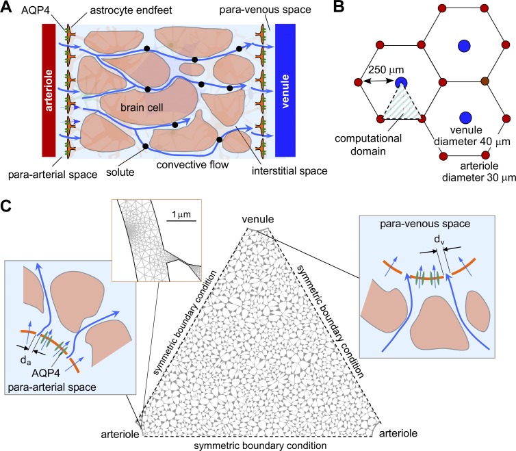
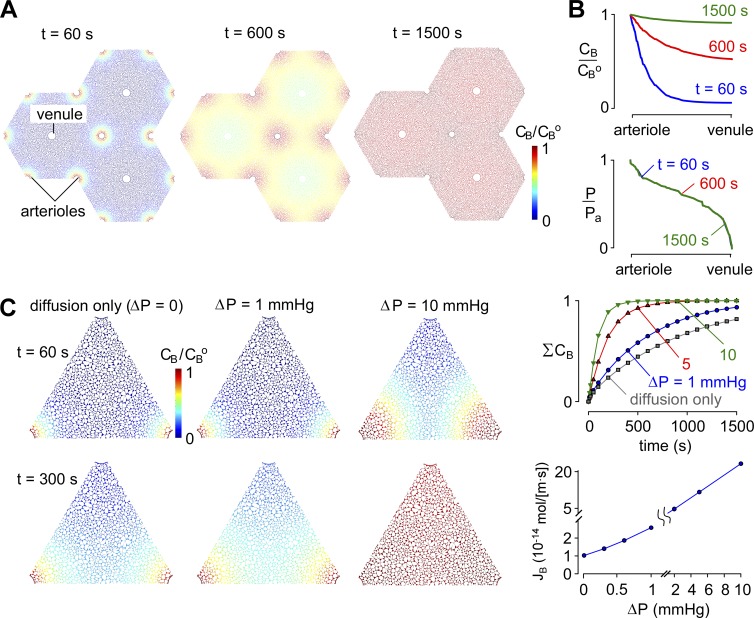
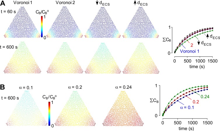
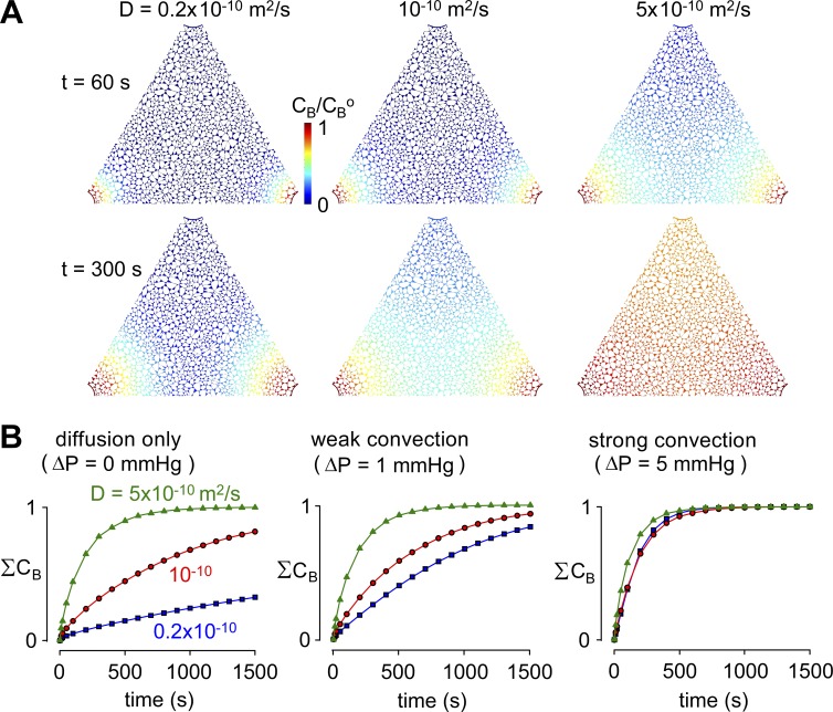
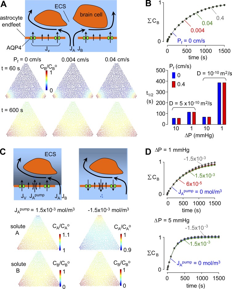
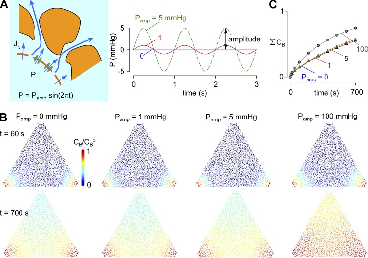
A "glymphatic system," which involves convective fluid transport from para-arterial to paravenous cerebrospinal fluid through brain extracellular space (ECS), has been proposed to account for solute clearance in brain, and aquaporin-4 water channels in astrocyte endfeet may have a role in this process. Here, we investigate the major predictions of the glymphatic mechanism by modeling diffusive and convective transport in brain ECS and by solving the Navier-Stokes and convection-diffusion equations, using realistic ECS geometry for short-range transport between para-arterial and paravenous spaces. Major model parameters include para-arterial and paravenous pressures, ECS volume fraction, solute diffusion coefficient, and astrocyte foot-process water permeability. The model predicts solute accumulation and clearance from the ECS after a step change in solute concentration in para-arterial fluid. The principal and robust conclusions of the model are as follows: (a) significant convective transport requires a sustained pressure difference of several mmHg between the para-arterial and paravenous fluid and is not affected by pulsatile pressure fluctuations; (b) astrocyte endfoot water permeability does not substantially alter the rate of convective transport in ECS as the resistance to flow across endfeet is far greater than in the gaps surrounding them; and (c) diffusion (without convection) in the ECS is adequate to account for experimental transport studies in brain parenchyma. Therefore, our modeling results do not support a physiologically important role for local parenchymal convective flow in solute transport through brain ECS.


Figures

Figure 1.
Figure 1.
Spatial model of convective fluid movement from para-arterial to paravenous spaces in brain ECS. (A) Schematic showing the major features of the proposed glymphatic mechanism, including convective fluid movement from para-arterial to paravenous spaces through brain extracellular (interstitial) space. (B) Hexagonal spatial arrangement of arterioles and venules in brain parenchyma, showing triangular computational domain. (C) Diagram of water and solute movement between the para-arterial space and ECS, and the ECS and the paravenous space. da and dv, gap distances between astrocyte endfeet in para-arterial and paravenous space.
Figure 2.
Figure 2.
General model predictions. (A) Pseudocolored images showing tracer concentration (CB/CBo) at time = 60, 600, and 1,500 s after a step increase in para-arterial tracer concentration for ΔP = 1 mmHg, Pf = 0.04 cm/s, and D = 10−10 m2/s. (B) CB/CBo and P/Po profiles from para-arterial to paravenous space at the indicated times. (C, left) Pseudocolored images showing tracer solute accumulation in ECS after a step increase in para-arterial tracer concentration for para-arterial to paravenous pressure differences ΔP of 0 mmHg (diffusion alone) or 1 or 10 mmHg. Parameters: Pf = 0.04 cm/s and D = 10−10 m2/s. (right, top) Kinetics of tracer solute accumulation in ECS for the indicated ΔP. (right, bottom) Steady-state tracer solute transfer from para-arterial to paravenous spaces for fixed concentration (CB = 0) at the paravenous space.
Figure 3.
Figure 3.
Pressure dependence of solute movement in brain ECS. (A) Influence of details of Voronoi cell geometry on model predictions. Four cell geometries were modeled: Voronoi 1 and 2, generated using different initial sets of random numbers with the same ECS width; Voronoi 3, with 25% reduced ECS width; and Voronoi 4, with 25% increased ECS width (each for the same ECS volume fraction). Images of CB/CBo shown in the triangular computational domain at t = 60 and 600 s (left) and the kinetics of spatially integrated CB in the ECS (ΣCB; right). (B) Influence of ECS volume fraction, α. Computations as in A for ΔP = 1 mmHg, for the indicated α. (left) Pseudocolored images as in A. (right) Kinetics of tracer solute accumulation in ECS for the indicated α.
Figure 4.
Figure 4.
Influence of tracer solute diffusion coefficient on solute movement in brain ECS. (A) Pseudocolored images showing tracer solute accumulation in ECS, as in Fig 3, for the indicated tracer solute diffusion coefficients, D, and for ΔP = 1 mmHg. Parameters: Pf = 0.04 cm/s and α = 0.2. (B) Kinetics of tracer solute accumulation in ECS for the indicated D and ΔP.
Figure 5.
Figure 5.
Influence of astrocyte endfoot water permeability, Pf, on solute movement in brain ECS. (A, top) Schematic showing hydrostatic and osmotic water transport across the astrocyte endfoot barrier and hydrostatic (advective) fluid movement in the gaps between endfeet. (bottom) Pseudocolored images showing tracer solute accumulation in ECS, as in Fig. 3, for Pf = 0, 0.004, and 0.04. For all computations in this figure, D = 10−10 m2/s and α = 0.2. (B, top) Kinetics of tracer solute accumulation in ECS for the indicated Pf. (bottom) Half-filling time for the indicated parameter sets. (C) Influence of active ion pumping to create an osmotic imbalance between the para-arterial space and ECS. (top) Schematic of possible effects of ion pumping into and out of the ECS across astrocyte endfeet, in which osmotically driven water transport across endfeet changes pressure in the ECS and hence the driving force for advective fluid movement from the para-arterial space into the ECS. (bottom) Pseudocolored images at time = 600 s for Pf = 0.04 cm/s showing accumulation of solutes A and B in ECS for the indicated active ion pumping flux, JApump = 1.5 × 10⁻³ mol/m3 (active pumping into the ECS) and JApump = −1.5 × 10⁻³ mol/m3 (pumping from the ECS). (D) Kinetics of tracer solute accumulation in ECS for ΔP = 1 and 5 mmHg.
Figure 6.
Figure 6.
Influence of pulsatile pressure in the para-arterial space on solute movement in brain ECS. (A, left) Schematic showing hydrostatic pressure driven in the para-arterial space water transport across the astrocyte endfoot barrier; (right) para-arterial pressure waveform of amplitude Pamp and frequency 1 Hz. (B) Pseudocolored images showing tracer solute accumulation in the ECS, as in Fig 3, for different Pamp. Parameters: Pf = 0.04 cm/s, D = 10−10 m2/s, and α = 0.2. (C) Kinetics of tracer solute accumulation in ECS for the indicated Pamp.

References

    1. Abbott N.J. 2004. Evidence for bulk flow of brain interstitial fluid: significance for physiology and pathology. Neurochem. Int. 45:545–552. 10.1016/j.neuint.2003.11.006 - DOI - PubMed
    1. Adams D.L., Piserchia V., Economides J.R., and Horton J.C.. 2015. Vascular supply of the cerebral cortex is specialized for cell layers but not columns. Cereb. Cortex. 25:3673–3681. 10.1093/cercor/bhu221 - DOI - PMC - PubMed
    1. Asgari M., de Zélicourt D., and Kurtcuoglu V.. 2015. How astrocyte networks may contribute to cerebral metabolite clearance. Sci. Rep. 5:15024 10.1038/srep15024 - DOI - PMC - PubMed
    1. Basser P.J. 1992. Interstitial pressure, volume, and flow during infusion into brain tissue. Microvasc. Res. 44:143–165. 10.1016/0026-2862(92)90077-3 - DOI - PubMed
    1. Bilston L.E., Fletcher D.F., Brodbelt A.R., and Stoodley M.A.. 2003. Arterial pulsation-driven cerebrospinal fluid flow in the perivascular space: a computational model. Comput. Methods Biomech. Biomed. Engin. 6:235–241. 10.1080/10255840310001606116 - DOI - PubMed

Publication types