. 2017 Jan;173(1):390-416.
doi: 10.1104/pp.16.01240. Epub 2016 Nov 8.

Membrane Proteomic Insights into the Physiology and Taxonomy of an Oleaginous Green Microalga

Affiliations

Membrane Proteomic Insights into the Physiology and Taxonomy of an Oleaginous Green Microalga

Adriana Garibay-Hernández et al. Plant Physiol. 2017 Jan.

Abstract

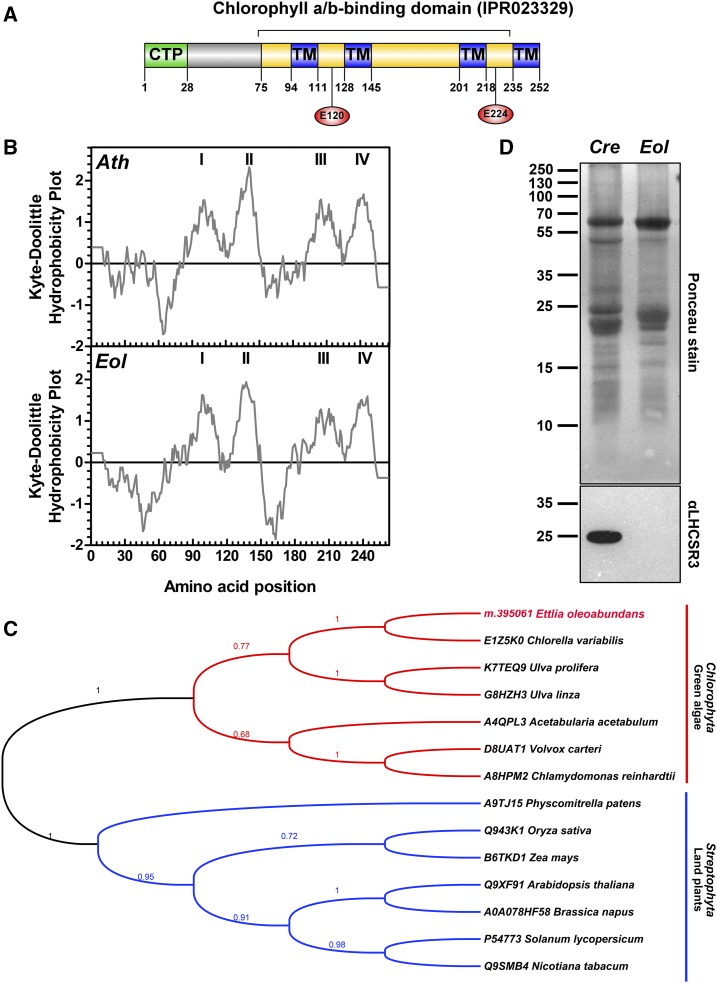
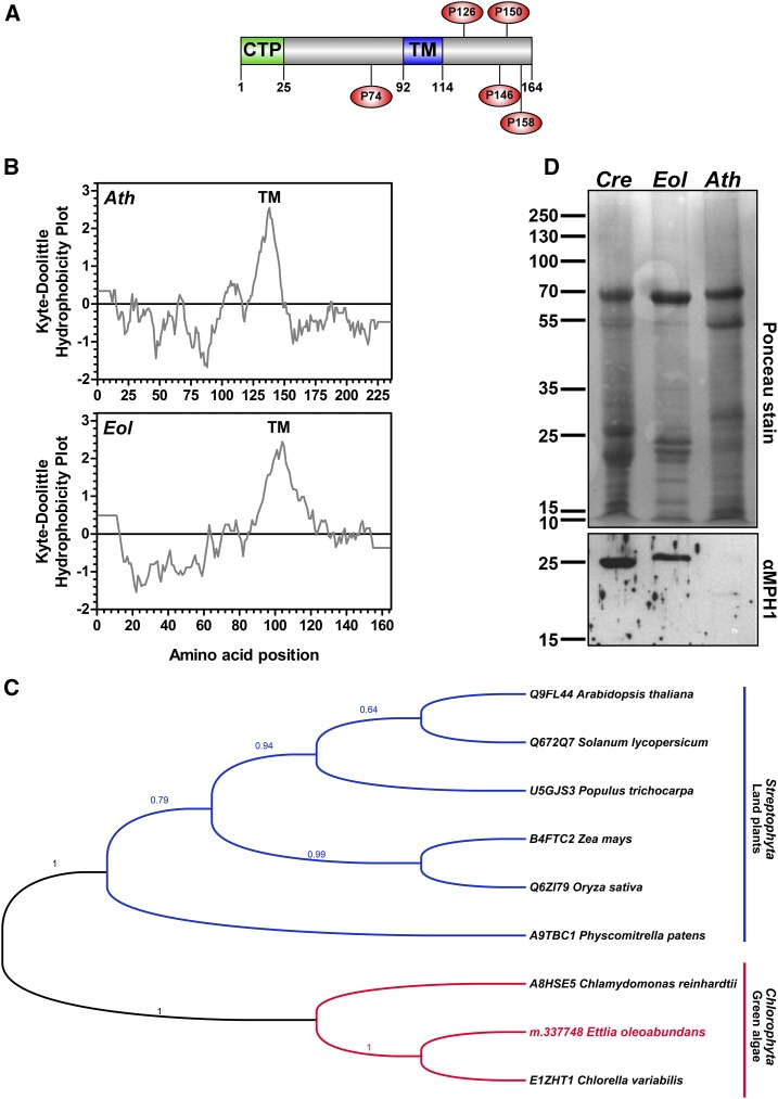
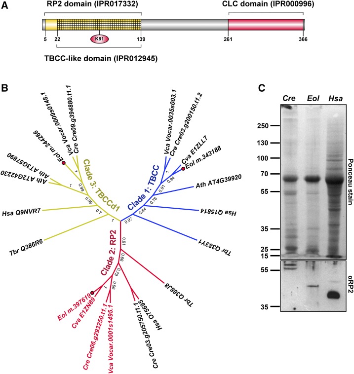
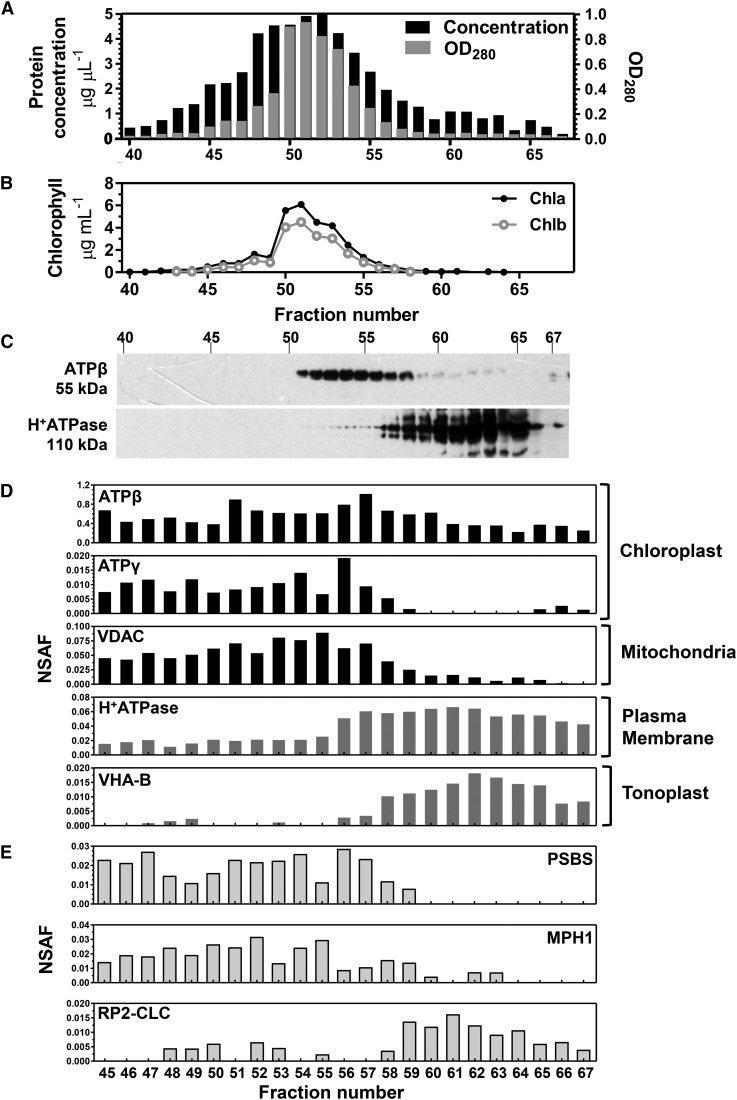
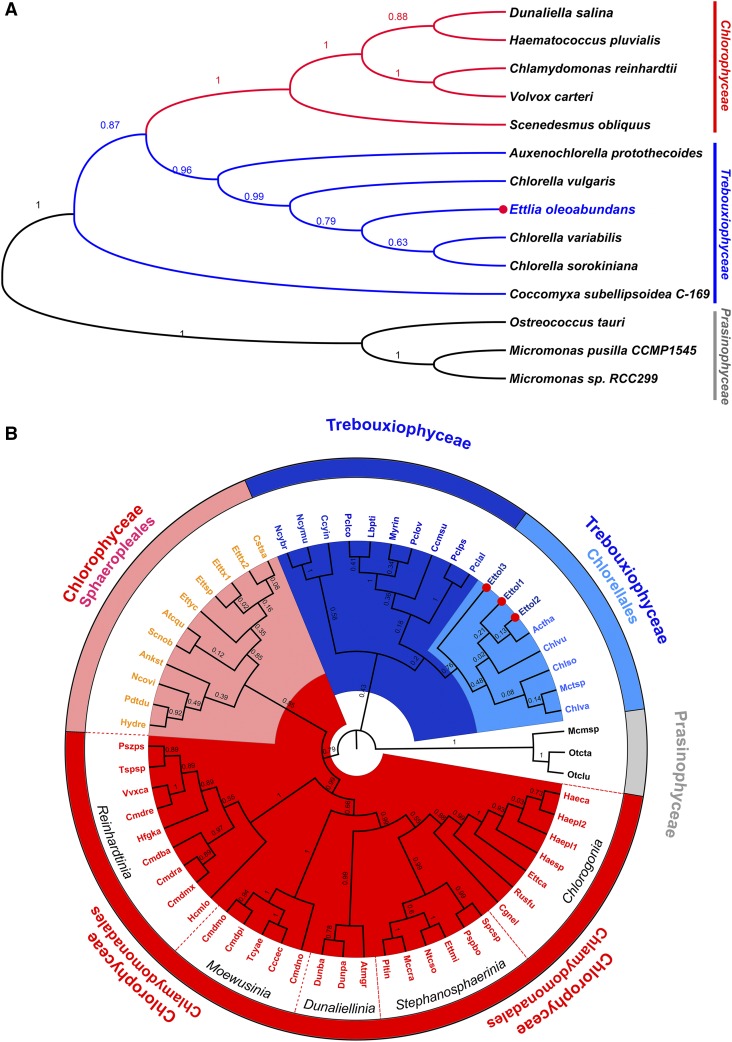
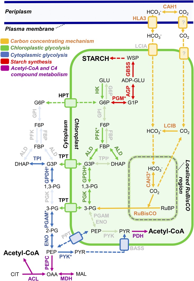
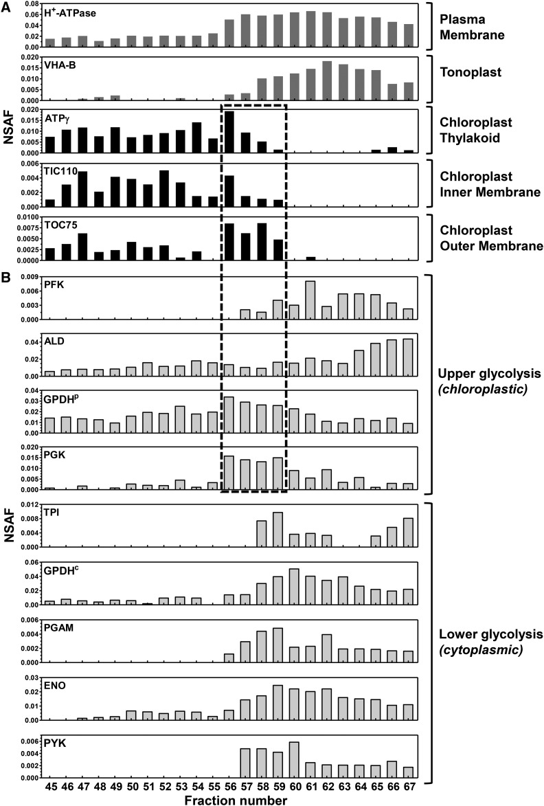
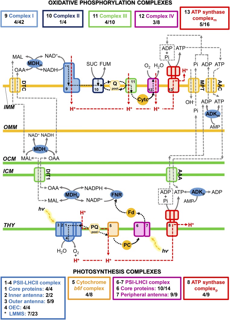
Ettlia oleoabundans is a nonsequenced oleaginous green microalga. Despite the significant biotechnological interest in producing value-added compounds from the acyl lipids of this microalga, a basic understanding of the physiology and biochemistry of oleaginous microalgae is lacking, especially under nitrogen deprivation conditions known to trigger lipid accumulation. Using an RNA sequencing-based proteomics approach together with manual annotation, we are able to provide, to our knowledge, the first membrane proteome of an oleaginous microalga. This approach allowed the identification of novel proteins in E. oleoabundans, including two photoprotection-related proteins, Photosystem II Subunit S and Maintenance of Photosystem II under High Light1, which were considered exclusive to higher photosynthetic organisms, as well as Retinitis Pigmentosa Type 2-Clathrin Light Chain, a membrane protein with a novel domain architecture. Free-flow zonal electrophoresis of microalgal membranes coupled to liquid chromatography-tandem mass spectrometry proved to be a useful technique for determining the intracellular location of proteins of interest. Carbon-flow compartmentalization in E. oleoabundans was modeled using this information. Molecular phylogenetic analyses of protein markers and 18S ribosomal DNA support the reclassification of E. oleoabundans within the trebouxiophycean microalgae, rather than with the Chlorophyceae class, in which it is currently classified, indicating that it may not be closely related to the model green alga Chlamydomonas reinhardtii A detailed survey of biological processes taking place in the membranes of nitrogen-deprived E. oleoabundans, including lipid metabolism, provides insights into the basic biology of this nonmodel organism.


Figures

Figure 1.
Figure 1.
Analysis of qE effector proteins (PSBS and LHCSR) in nitrogen-depleted E. oleoabundans. A, Protein architecture of EoPSBS. The identified protein domain signatures (InterPro; yellow), the predicted CTP (PredAlgo; green), and the transmembrane domains (TM; HMMTOP version 2.0; blue) are indicated. The conserved residues involved in the PSBS pH-sensing mechanism are shown (red circles). B, Hydrophobicity comparison of PSBS from E. oleoabundans (Eol) and Arabidopsis (Ath). Probable transmembrane domains (values greater than 0) are shown in the Kyte-Doolittle hydrophobicity plots (window size, 19). C, Phylogenetic analysis of PSBS homologs. Aligned sequences (Supplemental Fig. S3) were submitted for maximum likelihood (ML) analysis. The topology of the ML tree with the highest log likelihood (−3,122.6386) is shown. Bootstrap maximum likelihood (MLb) values are shown next to the branches. UniProtKB accession numbers for PSBS homologs are provided. D, Immunological detection of LHCSR in microsomes from E. oleoabundans and C. reinhardtii (Cre). The 12.5% (w/v) SDS-PAGE acrylamide gel was loaded with 20 μg of protein per lane.
Figure 2.
Figure 2.
MPH1 sequence analysis and identification in green microalgae membranes. A, Protein architecture of EoMPH1. The predicted CTP (PredAlgo; green) and transmembrane domain (TM; TMHMM version 2.0; blue) are indicated. Conserved Pro residues are shown (red circles). B, Hydrophobicity comparison of MPH1 from E. oleoabundans (Eol) and Arabidopsis (Ath). Probable transmembrane domains (values greater than 1.6) are shown in the Kyte-Doolittle hydrophobicity plots (window size, 19). C, Phylogenetic analysis of MPH1 homologs. Aligned sequences (Supplemental Fig. S4) were submitted for ML analysis. The topology of the ML tree with the highest log likelihood (−2,177.6951) is shown. MLb values are shown next to the branches. UniProtKB accession numbers for MPH1 homologs are provided. D, Immunological detection of MPH1 in microsomes from Arabidopsis (positive control; red box), E. oleoabundans, and C. reinhardtii (Cre). The 12.5% (w/v) SDS-PAGE acrylamide gel was loaded with 20 μg of protein per lane.
Figure 3.
Figure 3.
EoRP2-CLC is a membrane TBCC domain-containing protein with a novel domain architecture. A, Protein architecture of EoRP2-CLC. The identified InterPro signatures and the Lys residue (red circle) that may correspond to a homologous substitution of the key Arg residue for GAP activity are indicated. B, ML analysis of TBCC domain-containing proteins. Clade 1/TBCC is in blue, clade 2/RP2 is in red, and clade 3/TBCCd1 is in yellow. RP2-CLC domain architecture proteins are highlighted in red text. Amino acid sequences were aligned with webPRANK. The topology of the ML tree with the highest log likelihood (−8,226.6668) is shown. MLb values are shown next to the branches. Accession numbers are provided: UniProtKB (Hsa, Tbr, and Cva), Phytozome version 10 (Cre and Vca), and The Arabidopsis Information Resource (Ath). Ath, Arabidopsis; Cre, C. reinhardtii; Cva, C. variabilis; Hsa, H. sapiens; Eol, E. oleoabundans (red dots); Tbr, Trypanosoma brucei; Vca, Volvox carteri. C, Immunological detection of RP2-like proteins in microsomes from C. reinhardtii and E. oleoabundans. The 10% (w/v) SDS-PAGE acrylamide gel was loaded with 20 μg of protein per lane. Total protein extracts (10 μL) from human (Hsa) C2BBe1 cells (clone of Caco-2) were analyzed as a positive control, where the 40-kD RP2 human protein was identified.
Figure 4.
Figure 4.
Subcellular locations of novel proteins from E. oleoabundans via FFZE fractionation coupled to MS-based analysis. Microsomal membranes from nitrogen-deficient cultures were separated by FFZE. A, Protein profile of FFZE fractions. OD280, Optical density at 280 nm. B, Chlorophyll a and b concentrations in FFZE fractions. C, Immunological detection in the respective fractions of ATPβ (a chloroplast marker) and H+-ATPase (a plasma membrane marker). The 10% (w/v) SDS-PAGE acrylamide gel was loaded with 15 μg of protein per lane. The approximate molecular masses of the detected proteins are shown. D, Graphical representation of the normalized spectral count (NSAF values) of protein markers specific for subcellular compartments among the FFZE fractions. Individual FFZE fractions were analyzed by LC-MS/MS. The identification numbers for the surveyed protein markers are as follows: gi|416678 (ATPβ), m.392881 (ATPγ), m.378383 (VDAC), m.363780 (H+-ATPase), and m.395664 (VHA-B). E, Graphical representation of the NSAF values of PSBS, MPH1, and RP2-CLC among the FFZE fractions.
Figure 5.
Figure 5.
Molecular phylogenetic analysis of E. oleoabundans. A, ML analysis of concatenated nucleus-encoded (EF-1α and Actin), plastid-encoded (PSBA and RBCL), and mitochondria-encoded (COX1 and COX2) amino acid sequences (Supplemental Table S16). The topology of the ML tree with the highest log likelihood (−20,458.0427) is shown. B, ML analysis of 18S rDNA nucleotide sequences (Supplemental Table S17); Ettol2 corresponds to the sequence obtained in this work. The topology of the ML tree with the highest log likelihood (−12,721.9945) is shown. MLb values are shown next to the branches. E. oleoabundans sequences are highlighted (red dots). Green microalgae classes are denoted as follows: Chlorophyceae (red), Trebouxiophyceae (blue), and Prasinophyceae (gray; outgroup). Major taxa represented within these classes are denoted in B.
Figure 6.
Figure 6.
Carbon metabolism in nitrogen-deficient E. oleoabundans. Graphical representation is shown for the carbon metabolism proteins identified by LC-MS/MS in the membrane proteome of E. oleoabundans. All proteins were identified in FFZE fractions except hexokinase, which was identified exclusively in total microsome samples. Very-low-abundance proteins that were identified exclusively in FFZE fractions and not in total microsomes are highlighted with asterisks. Not identified proteins are shown in a clear gray color. Subcellular locations were predicted using PredAlgo together with experimental evidence available for the corresponding homologs. Identified proteins are described in Supplemental Table S4. Protein abbreviations are as follows: ACL, ATP-citrate synthase; AGP, Glc-1-P adenylyltransferase; ALD, aldolase; BASS, Bile Acid:Na+ Symporter, sodium/pyruvate cotransporter; CAH1, carbonic anhydrase, periplasmic; CAH3, carbonic anhydrase, chloroplastic; ENO, enolase; FBP, Fru-1,6-bisphosphatase; G6P, Glc-6-phosphatase; GBSS, granule-bound starch synthase; GPDHc, glyceraldehyde-3-phosphate dehydrogenase, cytosolic; GPDHp, glyceraldehyde-3-phosphate dehydrogenase A, chloroplastic; GPI, Glc-6-P isomerase; HLA3, probable inorganic carbon transporter HLA3; HK, hexokinase; HPT, UhpC-type hexose phosphate translocator; LCIA, putative inorganic carbon transporter LCIA; LCIB, LCIB family protein; MDH, malate dehydrogenase, cytoplasmic; PDH, pyruvate dehydrogenase; PEPC, phosphoenolpyruvate carboxylase; PFK, phosphofructokinase; PGAM, phosphoglycerate mutase; PGK, phosphoglycerate kinase; PGM, phosphoglucomutase; PPT, phosphoenolpyruvate/phosphate translocator; PYK, pyruvate kinase; TPI, triose phosphate isomerase; TPT, triose phosphate/phosphate translocator; RuBisCO, ribulose-1,5-biphosphate carboxylase. Compound abbreviations are as follows: ADP-GLU, ADP-Glc; CIT, citrate; DHAP, dihydroxyacetone phosphate; FBP, Fru-1,6-bisphosphate; F6P, Fru-6-P; G3P, glyceraldehyde-3-phosphate; GLU, Glc; G1P, Glc-1-P; G6P, Glc-6-P; MAL, malate; OAA, oxaloacetate; PEP, phosphoenolpyruvate; 1,3-PG, 1,3-bisphosphoglycerate; 2-PG, 2-phosphoglycerate; 3-PG, 3-phosphoglycerate; PYR, pyruvate; RuBP, ribulose-1,5-biphosphate; WSP, water-soluble polysaccharide.
Figure 7.
Figure 7.
FFZE profiles of glycolytic enzymes suggest their targeting to multiple cellular locations. Individual FFZE fractions were analyzed by LC-MS/MS and surveyed for proteins of interest. A, Graphical representation of NSAF values of protein markers specific for subcellular compartments among the FFZE fractions. The surveyed compartment markers are as follows: m.363780 (H+-ATPase), m.392881 (ATPγ), m.227792 (TIC110), and m.134654 (TOC75). B, Graphical representation of NSAF values of glycolytic enzymes among the FFZE fractions. According to their predicted cellular locations, proteins are grouped into upper and lower glycolytic pathway enzymes. These proteins are described in Supplemental Table S4. FFZE fractions enriched with the outer chloroplast membrane are enclosed in the box (fractions 56–59).
Figure 8.
Figure 8.
Photosynthesis and oxidative phosphorylation in the membrane proteome of nitrogen-deficient E. oleoabundans. Graphical representation is shown for the proteins identified by LC-MS/MS in the membrane proteome of E. oleoabundans involved in energy conversion and homeostasis. Identified proteins are described in Supplemental Table S1 (photosynthesis), Supplemental Table S2 (oxidative phosphorylation), and Supplemental Table S3 (energy and reducing power homeostasis). The number of proteins detected in E. oleoabundans from each of the complexes of the chloroplastic and mitochondrial electron transfer chains is shown in colored boxes and compared with the number of proteins identified in the complexes of the model alga C. reinhardtii. In complex IV, CrCOX2 is considered as a nonsplit subunit. In the E. oleoabundans ATP synthase mitochondrial complex, the N- and C-terminal peptides of subunit d are considered as a unique nonsplit protein. Black dashed lines indicate electron transfer, and red dashed lines indicate proton translocation. C, Cytoplasmic; M, mitochondrial; P, chloroplastic. Asterisks indicate proteins associated with any part of the PSII-LHCII supercomplex. AAA, ADP/ATP carrier protein, chloroplastic; AAC, ADP/ATP carrier protein, mitochondrial; ADK, adenylate kinase; Cytc, cytochrome c; DIT1, dicarboxylate transporter 1, chloroplastic; DTC, mitochondrial dicarboxylate/tricarboxylate carrier; Fd, ferredoxin; FNR, ferredoxin-NADP reductase; FUM, fumarate; ICM, inner chloroplast membrane; IMM, inner mitochondrial membrane; LHCI, light-harvesting complex of PSI; LHCII, light-harvesting complex of PSII; LMMS, low-molecular-mass subunits; MAL, malate; MDH, malate dehydrogenase; MPT, mitochondrial phosphate carrier protein; OAA, oxaloacetate; OCM, outer chloroplast membrane; OEC, oxygen-evolving complex; OMM, outer mitochondrial membrane; PC, plastocyanin; Pi, inorganic phosphate; PQ, plastoquinone; Q, ubiquinone; SUC, succinate; THY, thylakoid membrane.

Similar articles

Cited by

References

    1. Ajjawi I, Coku A, Froehlich JE, Yang Y, Osteryoung KW, Benning C, Last RL (2011) A J-like protein influences fatty acid composition of chloroplast lipids in Arabidopsis. PLoS ONE 6: e25368. - PMC - PubMed
    1. Alboresi A, Gerotto C, Giacometti GM, Bassi R, Morosinotto T (2010) Physcomitrella patens mutants affected on heat dissipation clarify the evolution of photoprotection mechanisms upon land colonization. Proc Natl Acad Sci USA 107: 11128–11133 - PMC - PubMed
    1. Arnon DI. (1949) Copper enzymes in isolated chloroplasts: polyphenoloxidases in Beta vulgaris. Plant Physiol 24: 1–15 - PMC - PubMed
    1. Arredondo-Vega BO, Band CJ, Vazquez-Duhalt R (1995) Biochemical composition of Neochloris oleoabundans adapted to marine medium. Cytobios 83: 201–205
    1. Atteia A, Adrait A, Brugière S, Tardif M, van Lis R, Deusch O, Dagan T, Kuhn L, Gontero B, Martin W, et al. (2009) A proteomic survey of Chlamydomonas reinhardtii mitochondria sheds new light on the metabolic plasticity of the organelle and on the nature of the α-proteobacterial mitochondrial ancestor. Mol Biol Evol 26: 1533–1548 - PubMed

Publication types

MeSH terms

LinkOut - more resources