. 2017 Jan;173(1):728-741.
doi: 10.1104/pp.16.01253. Epub 2016 Nov 8.

Extracellular Vesicles Isolated from the Leaf Apoplast Carry Stress-Response Proteins

Affiliations

Extracellular Vesicles Isolated from the Leaf Apoplast Carry Stress-Response Proteins

Brian D Rutter et al. Plant Physiol. 2017 Jan.

Abstract

Exosomes are extracellular vesicles (EVs) that play a central role in intercellular signaling in mammals by transporting proteins and small RNAs. Plants are also known to produce EVs, particularly in response to pathogen infection. The contents of plant EVs have not been analyzed, however, and their function is unknown. Here, we describe a method for purifying EVs from the apoplastic fluids of Arabidopsis (Arabidopsis thaliana) leaves. Proteomic analyses of these EVs revealed that they are highly enriched in proteins involved in biotic and abiotic stress responses. Consistent with this finding, EV secretion was enhanced in plants infected with Pseudomonas syringae and in response to treatment with salicylic acid. These findings suggest that EVs may represent an important component of plant immune responses.


Figures

Figure 1.
Figure 1.
The apoplastic wash from Arabidopsis leaves contains vesicle-like structures. A and B, Negative staining and TEM of the P40 and P100 fractions derived from the apoplastic wash. C and D, Light-scattering charts showing peak intensities and percentages of differently sized particles in the P40 and P100 fractions. The experiments were repeated a minimum of three times with similar results.
Figure 2.
Figure 2.
Arabidopsis EVs contain the syntaxin PEN1. A, Confocal microscopy images (inverted) of the P40 and P100 fractions derived from the apoplastic wash of nontransgenic (Columbia-0 [Col-0]) and GFP-PEN1 transgenic plants. Bars = 10 µm. B, Quantification of fluorescent foci from confocal microscopy images. Letters signify which values are significantly different from each other based on a two-tailed unpaired Student's t test (P < 0.05). C, Immunoblots of the P40 and P100 fractions confirm that GFP-PEN1 is present only in the P40 fraction of transgenic GFP-PEN1 plants. The lysate lane indicates whole-leaf protein extracts. D, GFP-PEN1 expressed under the control of a native promoter (NP) also accumulates in the P40 fraction. E, Detergent treatment removes GFP-PEN1 from the P40 fraction. P40 fractions derived from GFP-PEN1 plants were treated with buffer alone or buffer plus TX100 followed by recentrifugation. Treatment with TX100 removed GFP-PEN1 from the pellet. F, GFP-PEN1 in the P40 fraction is protected from trypsin degradation in the absence of detergent. All experiments were repeated a minimum of three times with similar results. Error bars in B indicate sd; n = 4; P < 0.05 using a two-tailed unpaired Student’s t test.
Figure 3.
Figure 3.
Apoplastic EVs are enriched for PEN1. A, Transgenic lines constitutively expressing the indicated endomembrane and PM markers were subjected to EV isolation, and the P40 fractions were analyzed by immunoblot. The lysate blot indicates whole-leaf protein extracts. Only GFP-PEN1 was readily detectable in the P40 fraction. B, Quantification of marker proteins in the P40 fraction. Band intensities were quantified, and the ratio between the P40 and lysate bands was calculated for each protein. PATL1, an EV-localized protein that we identified by mass spectrometry (Supplemental Table S1), was used to normalize for EV concentrations in the P40 fractions. The specificity of the anti-PATL1 antibody is shown in Supplemental Figure S1. This experiment was repeated a minimum of three times with similar results.
Figure 4.
Figure 4.
Density gradient purification of EVs. A, Immunoblot detection of GFP-PEN1 in fractions from the iodixanol density gradient. Fraction 1 consisted of 3.5 mL, fraction 2 was 1.7 mL, fractions 3 to 7 were 0.6 mL each, and fractions 8 to 12 were 0.7 mL each. B, Light-scattering charts showing peak intensities for pooled fractions 2 to 4. C, Confocal microscopy images (inverted) of Tris-HCl, pH 7.5, control (left) and pooled fractions 2 to 4 resuspended in Tris-HCl, pH 7.5 (right). Bars = 10 µm. D, Quantification of confocal images in C. Letters signify values that are significantly different from each other based on a two-tailed unpaired Student's t test (P < 0.0001). E, Negative staining and TEM of fractions 2 to 4. Bar = 10 µm. F, Cryo-electron microscopy of fractions 2 to 4. Note the lipid bilayer in the bottom right vesicle. Bar = 100 nm. All experiments were repeated at least two times with similar results. Error bars indicate sd; n = 3; P < << 0.001 using a two-tailed unpaired Student’s t test.
Figure 5.
Figure 5.
Plant EVs are enriched for stress-response proteins. The entire Arabidopsis proteome (top), the EV proteome from uninfected plants (bottom left), and the EV proteome from P. syringae-infected plants (bottom right) were categorized based on GO terms through The Arabidopsis Information Resource Web site (www.arabidopsis.org). These analyses are based on the data given in Supplemental Table S1 and are derived from two biological replicates.
Figure 6.
Figure 6.
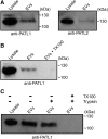
Plant EVs are associated with PATL1 and PATL2. A, Immunoblots of the P40 fraction and whole-leaf protein extracts using PATL1 and PATL2 antibodies. B, Detergent treatment removes PATL1 from the P40 fraction. P40 fractions derived from Col-0 plants were treated with buffer alone or buffer plus TX100. Following recentrifugation, the sample treated with TX100 contained much less PATL1. C, PATL1 in the P40 fraction is protected from trypsin degradation in the absence of detergent. All experiments were repeated a minimum of two times with similar results.
Figure 7.
Figure 7.
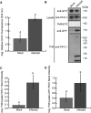
EV secretion is enhanced during P. syringae infection. A, Total membrane content in the P40 fraction increases over 2-fold following spray inoculation with a virulent P. syringae strain. Col-0 Arabidopsis plants were sprayed with either P. syringae DC3000 (pVPS61:empty) at an optical density at 600 nm of 0.2 or a control solution lacking bacteria. EVs were isolated 3 d after the initial infection from three sets of plants per treatment. Each sample of EVs was stained with 3,3′-dihexyloxacarbocyanine iodide (DiOC6), washed, and repelleted. Fluorescence intensity was quantified (error bars indicate sd; n = 3; P < < 0.001 using a two-tailed unpaired Student’s t test). B to D, PATL1 and GFP-PEN1 contents in the P40 fraction increase over 2-fold following P. syringae infection. B, Representative P40 fractions for both treatments, as well as samples of total cellular lysate, were immunoblotted for full-length PATL1. The levels of PATL1 are higher in the P40 fraction from infected plants compared with the P40 fraction from mock-infected plants, as indicated by a more intense band around 130 kD and the appearance of strong degradation products around 20 and 10 kD. C and D, Quantification of PATL1 and GFP-PEN1 band intensities from B. The experiment was repeated three times with similar results. The results of only one experiment are shown.
Figure 8.
Figure 8.
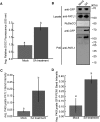
EV secretion is enhanced after SA treatment. A, Total membrane content in the P40 fraction increases over 2-fold following treatment with SA. Col-0 Arabidopsis plants were sprayed with either 2 mm SA or a control solution lacking SA. EVs were isolated 12 h after spraying from three sets of plants per treatment. Each sample of EVs was stained with DiOC6, washed, and repelleted. Fluorescence intensity was quantified (error bars indicate sd; n = 3; P < < 0.001 using a two-tailed unpaired Student’s t test). B to D, PATL1 and GFP-PEN1 levels in the P40 fraction increase over 3-fold following SA treatment. B, Representative P40 fractions, as well as samples of total cellular lysate, were immunoblotted for full-length PATL1. The levels of PATL1 are higher in the P40 fraction from SA-treated plants, as indicated by a more intense band around 130 kD. C and D, Quantification of PATL1 and GFP-PEN1 band intensities from B. The experiment was repeated three times with similar results. The results of only one experiment are shown.

Similar articles

Cited by

References

    1. Admyre C, Johansson SM, Qazi KR, Filén JJ, Lahesmaa R, Norman M, Neve EP, Scheynius A, Gabrielsson S (2007) Exosomes with immune modulatory features are present in human breast milk. J Immunol 179: 1969–1978 - PubMed
    1. Agrawal GK, Jwa NS, Lebrun MH, Job D, Rakwal R (2010) Plant secretome: unlocking secrets of the secreted proteins. Proteomics 10: 799–827 - PubMed
    1. Akers JC, Gonda D, Kim R, Carter BS, Chen CC (2013) Biogenesis of extracellular vesicles (EV): exosomes, microvesicles, retrovirus-like vesicles, and apoptotic bodies. J Neurooncol 113: 1–11 - PMC - PubMed
    1. Alexandersson E, Saalbach G, Larsson C, Kjellbom P (2004) Arabidopsis plasma membrane proteomics identifies components of transport, signal transduction and membrane trafficking. Plant Cell Physiol 45: 1543–1556 - PubMed
    1. Aline F, Bout D, Amigorena S, Roingeard P, Dimier-Poisson I (2004) Toxoplasma gondii antigen-pulsed-dendritic cell-derived exosomes induce a protective immune response against T. gondii infection. Infect Immun 72: 4127–4137 - PMC - PubMed

MeSH terms