. 2017 Apr;28(4):1162-1174.
doi: 10.1681/ASN.2015111266. Epub 2016 Nov 11.

Chronic Hyperphosphatemia and Vascular Calcification Are Reduced by Stable Delivery of Soluble Klotho

Affiliations

Chronic Hyperphosphatemia and Vascular Calcification Are Reduced by Stable Delivery of Soluble Klotho

Julia M Hum et al. J Am Soc Nephrol. 2017 Apr.

Abstract

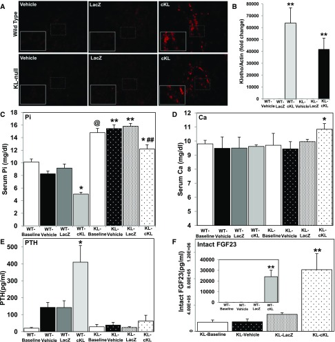
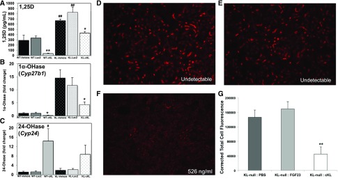
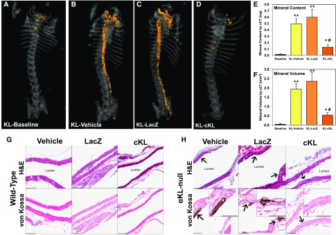
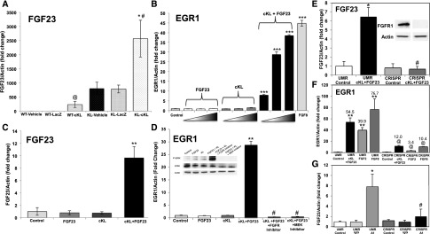
αKlotho (αKL) regulates mineral metabolism, and diseases associated with αKL deficiency are characterized by hyperphosphatemia and vascular calcification (VC). αKL is expressed as a membrane-bound protein (mKL) and recognized as the coreceptor for fibroblast growth factor-23 (FGF23) and a circulating soluble form (cKL) created by endoproteolytic cleavage of mKL. The functions of cKL with regard to phosphate metabolism are unclear. We tested the ability of cKL to regulate pathways and phenotypes associated with hyperphosphatemia in a mouse model of CKD-mineral bone disorder and αKL-null mice. Stable delivery of adeno-associated virus (AAV) expressing cKL to diabetic endothelial nitric oxide synthase-deficient mice or αKL-null mice reduced serum phosphate levels. Acute injection of recombinant cKL downregulated the renal sodium-phosphate cotransporter Npt2a in αKL-null mice supporting direct actions of cKL in the absence of mKL. αKL-null mice with sustained AAV-cKL expression had a 74%-78% reduction in aorta mineral content and a 72%-77% reduction in mineral volume compared with control-treated counterparts (P<0.01). Treatment of UMR-106 osteoblastic cells with cKL + FGF23 increased the phosphorylation of extracellular signal-regulated kinase 1/2 and induced Fgf23 expression. CRISPR/Cas9-mediated deletion of fibroblast growth factor receptor 1 (FGFR1) or pretreatment with inhibitors of mitogen-activated kinase kinase 1 or FGFR ablated these responses. In summary, sustained cKL treatment reduced hyperphosphatemia in a mouse model of CKD-mineral bone disorder, and it reduced hyperphosphatemia and prevented VC in mice without endogenous αKL. Furthermore, cKL stimulated Fgf23 in an FGFR1-dependent manner in bone cells. Collectively, these findings indicate that cKL has mKL-independent activity and suggest the potential for enhancing cKL activity in diseases of hyperphosphatemia with associated VC.

Keywords: FGF23; alpha-klotho; bone; db/db-eNOS; hyperphosphatemia; osteocyte.


Figures

Figure 1.
Figure 1.
cKL rescues hyperphosphatemia in a model of diabetic renal failure. (A) Representative differences in histopathologic changes as shown by three staining methods, H&E, periodic acid–Schiff (PAS), and Masson trichrome (MTS), in kidneys from (i–iii) control lean (db/dm) mice and (iv–vi) db/db-eNOS−/− LacZ mice. There is prominent presence of tubular protein (arrowheads and inset in iv) and dilated tubules (arrows in iv) in the H&E-stained sections from db/db-eNOS−/− LacZ mice that was absent in control lean mice (inset i). In db/db-eNOS−/− LacZ mice there was notable mesangial matrix deposition highlighted by PAS staining (arrows and inset v) that obliterates normal capillary loops and cellularity in the glomeruli found in control lean mice (arrows and inset ii). Marked interstitial fibrosis highlighted in blue by the MTS stain (stars in vi) and prominent glomerular fibrosis (arrows and inset in vi) with occasional glomerular sclerosis (arrow head in vi) in db/db-eNOS−/− LacZ mice and absent in control lean mice (inset iii). (B) Male and female body weights (grams) from baseline and 2, 4, and 6 weeks post-treatment. All db/db-eNOS−/− mice, regardless of treatment, were significantly heavier than age–matched lean control mice. *P<0.01. (C) Blood glucose was elevated above baseline lean controls in male and female db/db-eNOS−/− mice, regardless of treatment. *P<0.01. (D) Urine ACR was significantly elevated in male and female db/db-eNOS−/− mice compared with age–matched lean controls. ϕP<0.001. (E) Serum calcium was reduced in AAV-cKL–treated female db/db-eNOS−/− mice at 2, 4, and 6 weeks postinjection compared with AAV-LacZ–treated female db/db-eNOS−/− mice and age–matched lean controls. By 6 weeks, serum calcium had significantly risen in AAV-LacZ female db/db-eNOS−/− mice compared with lean controls. *P<0.05 (age–matched lean controls); @P<0.05 (AAV-LacZ–treated female db/db-eNOS−/− mice). (F) Serum Klotho levels in db/db-eNOS−/− male and female mice were significantly increased with AAV-cKL treatment 6 weeks postinjection. *P<0.05 versus all AAV-LacZ and lean mice; **P<0.01 versus all AAV-LacZ and lean mice. (G) Intact FGF23 was significantly elevated in db/db-eNOS−/− male and female mice with AAV-cKL treatment 4 weeks postinjection and remained elevated at 6 weeks. *P<0.05. (H) Serum phosphate was significantly reduced in female db/db-eNOS−/− mice by 4 and 6 weeks post–AAV-cKL injection compared with female db/db-eNOS−/− mice injected with AAV-LacZ and lean controls. Female db/db-eNOS−/− mice treated with AAV-LacZ had significantly elevated serum phosphate by 6 weeks after treatment. *P<0.05 (lean controls); @P<0.05 (AAV-LacZ mice); #P<0.05 versus 2 and 4 weeks.
Figure 2.
Figure 2.
Biochemical and endocrine effects of AAV-cKL in WT and Klotho-null mice. (A) Liver sections from WT and αKL-null mice were examined for αKL expression by immunofluorescence staining. αKL protein was only observed in mice delivered AAV-cKL, and staining (red) localized to cells near hepatic portal venules. (B) WT and αKL-null livers were assessed for cKL mRNA expression. WT mice and αKL-null mice treated with AAV-cKL had highly expressed cKL mRNA (approximately 60,000- and 40,000-fold increases, respectively). There was no significant difference between AAV-cKL–treated mice. **P<0.01. (C) αKL-null mice were hyperphosphatemic compared with WT mice at baseline and across treatment groups. Compared with like genotype controls (vehicle or LacZ), serum phosphate (Pi) was reduced in WT and Klotho mice treated with cKL. *P<0.01; **P<0.001 (across treatment groups); ##P<0.01 (across treatment groups); @P<0.001 (baseline). (D) Within genotypic groups, serum calcium (Ca) was unaffected by 4 weeks of cKL treatment. However, a slight elevation in serum calcium in Klotho-cKL was significant compared with WT-cKL. *P<0.05 versus WT-cKL. (E) cKL induced hyperparathyroidism in WT mice, but Klotho mice exhibited sustained hypoparathyroidism, regardless of treatment. *P<0.05. (F, inset) Intact FGF23 was significantly increased in WT mice treated with cKL. **P<0.01 versus baseline, vehicle, and AAV-LacZ. (F) KL mice had baseline elevated intact FGF23 versus WT mice at baseline and displayed a further increase with cKL treatment. **P<0.01 versus vehicle and LacZ; @P<0.05 (baseline).
Figure 3.
Figure 3.
cKL influences renal gene expression. (A) Serum 1,25D concentrations were tested in WT and αKL-null mice after 4 weeks of treatment. Sustained delivery of AAV-cKL in WT mice reduced 1,25D. αKL-null mice exhibit elevated 1,25D; AAV-cKL reduced 1,25D. *P<0.05; **P<0.01; ##P<0.01. (B) Delivery of AAV-cKL reduced renal expression of 1α-OHase (Cyp27b1) in both WT and αKL-null mice. *P<0.05. (C) Renal expression of 24-OHase (Cyp24a1) was increased in WT-cKL mice, whereas in KL-cKL mice, there was a trend for 24-OHase elevation, which was not statistically significant. #P<0.05. (D) αKL-null mice were injected intravenously with PBS, FGF23 (1 μg/g body wt), or cKL (1 μg/g body wt) for 1 hour and then, analyzed for kidney Npt2a expression by immunofluorescence. (D) Vehicle (cKL undetectable) and (E) FGF23-injected mice (cKL undetectable) had similar levels of Npt2a. (F) After injection with cKL, Npt2a expression was reduced. The concentration represents the serum cKL levels after injection, assuring cKL delivery (cKL range =214–526 ng/ml). (G) Quantitative analysis showed a significant reduction of Npt2a expression in mice treated with cKL versus controls. **P<0.01.
Figure 4.
Figure 4.
cKL prevents aortic calcification. (A) Representative μCT images of aortic calcification (orange) from Klotho mice from baseline (4 weeks of age) as well as (B) vehicle, (C) LacZ, and (D) cKL groups (treated from 4 weeks of age for 4 additional weeks). cKL administration was associated with a visually marked reduction in aortic mineralization versus Klotho-vehicle and Klotho-LacZ. (E) Mineral content and (F) mineral volume of whole aortas were quantified and determined to be significantly elevated versus all groups; however, in cKL-treated mice, mineral content and volume were significantly reduced versus Klotho-vehicle and Klotho-LacZ. Longitudinal sections of aorta from treated mice were examined by histology. *P<0.05 versus baseline; **P<0.01 versus baseline; #P<0.01 versus vehicle and AAV-LacZ treated. (G) Compared with WT mice, (H) αKL-null mice showed multifocal disruption or expansion of the aortic elastic lamina (arrows). αKL-null mice aortas from vehicle and AAV-LacZ–treated mice were positively stained with von Kossa (dark patches are indicative of mineralization), but <50% of such lesions from mice administered cKL stained positive (insets). Scale bar, 100 μm.
Figure 5.
Figure 5.
cKL signals in bone cells via FGFR1. (A) FGF23 mRNA was increased in femurs of WT mice treated with AAV-cKL. αKL-null mice had elevated bone FGF23 expression compared with WT mice, which was further increased in αKL-null mice treated with AAV-cKL. *P<0.01 versus same genotype controls; #P<0.01 versus WT mice; @P<0.01 versus controls. (B) UMR-106 cells treated with cKL or FGF23 alone showed no effects; however, a combination of cKL + FGF23 increased expression of EGR1 mRNA. The positive control FGF8 increased EGR1 mRNA. ***P<0.001 versus control (untreated). (C) FGF23 mRNA was significantly increased in UMR-106 cells administered cKL + FGF23. **P<0.01 versus all other treatments. (D) EGR1 mRNA increased in response to cKL + FGF23 and was dependent on functional FGFR signaling and MEK activity, because pretreatment with either an FGFR or MEK inhibitor blocked EGR1 mRNA production as well as p-ERK expression by immunoblot (inset). **P<0.01 versus control and FGF23 or cKL alone; #P<0.001 versus cKL + FGF23. (E) A novel UMR-106 cell line with deletion of FGFR1 was generated via CRISPR-Cas. Immunoblots of lysates from the parent UMR-106 line (UMR) and the CRISPR-targeted line (CRISPR) assured ablated FGFR1 protein (inset). cKL + FGF23 increased FGF23 mRNA expression in the UMR cells, but FGF23 expression was not different from UMR control in the CRISPR line. P value was not significant versus UMR control. *P<0.05 versus UMR control (untreated); #P<0.001 versus UMR cKL + FGF23. (F) EGR1 mRNA expression was increased in response to FGF2, FGF8, and cKL + FGF23 in UMR cells. This response was significantly blunted in the CRISPR cells. **P<0.001 versus UMR control; @P<0.05 versus UMR respective treatment. (G) Administration of an FGFR1c agonist antibody increased FGF23 mRNA expression in the parent UMR cells but failed to upregulate FGF23 in CRISPR cells. *P<0.05 versus UMR control; #P<0.05 versus UMR A1.

References

    1. Fliser D, Kollerits B, Neyer U, Ankerst DP, Lhotta K, Lingenhel A, Ritz E, Kronenberg F, Kuen E, König P, Kraatz G, Mann JF, Müller GA, Köhler H, Riegler P, Riegler P: Fibroblast growth factor 23 (FGF23) predicts progression of chronic kidney disease: The Mild to Moderate Kidney Disease (MMKD) Study. J Am Soc Nephrol 18: 2600–2608, 2007 - PubMed
    1. Mirza MA, Larsson A, Melhus H, Lind L, Larsson TE: Serum intact FGF23 associate with left ventricular mass, hypertrophy and geometry in an elderly population. Atherosclerosis 207: 546–551, 2009 - PubMed
    1. Gutiérrez OM, Mannstadt M, Isakova T, Rauh-Hain JA, Tamez H, Shah A, Smith K, Lee H, Thadhani R, Jüppner H, Wolf M: Fibroblast growth factor 23 and mortality among patients undergoing hemodialysis. N Engl J Med 359: 584–592, 2008 - PMC - PubMed
    1. Scialla JJ, Xie H, Rahman M, Anderson AH, Isakova T, Ojo A, Zhang X, Nessel L, Hamano T, Grunwald JE, Raj DS, Yang W, He J, Lash JP, Go AS, Kusek JW, Feldman H, Wolf M; Chronic Renal Insufficiency Cohort (CRIC) Study Investigators : Fibroblast growth factor-23 and cardiovascular events in CKD. J Am Soc Nephrol 25: 349–360, 2014 - PMC - PubMed
    1. Ix JH, Katz R, Kestenbaum BR, de Boer IH, Chonchol M, Mukamal KJ, Rifkin D, Siscovick DS, Sarnak MJ, Shlipak MG: Fibroblast growth factor-23 and death, heart failure, and cardiovascular events in community-living individuals: CHS (Cardiovascular Health Study). J Am Coll Cardiol 60: 200–207, 2012 - PMC - PubMed