. 2017 Feb:36:43-52.
doi: 10.1016/j.jocn.2016.10.012. Epub 2016 Nov 9.

Dysregulation of PINCH signaling in mesial temporal epilepsy

Affiliations

Dysregulation of PINCH signaling in mesial temporal epilepsy

Charles Liu et al. J Clin Neurosci. 2017 Feb.

Abstract

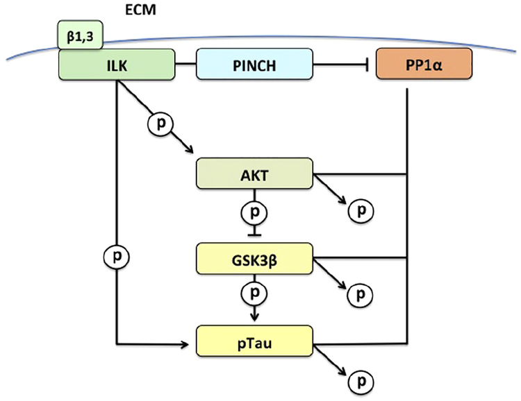
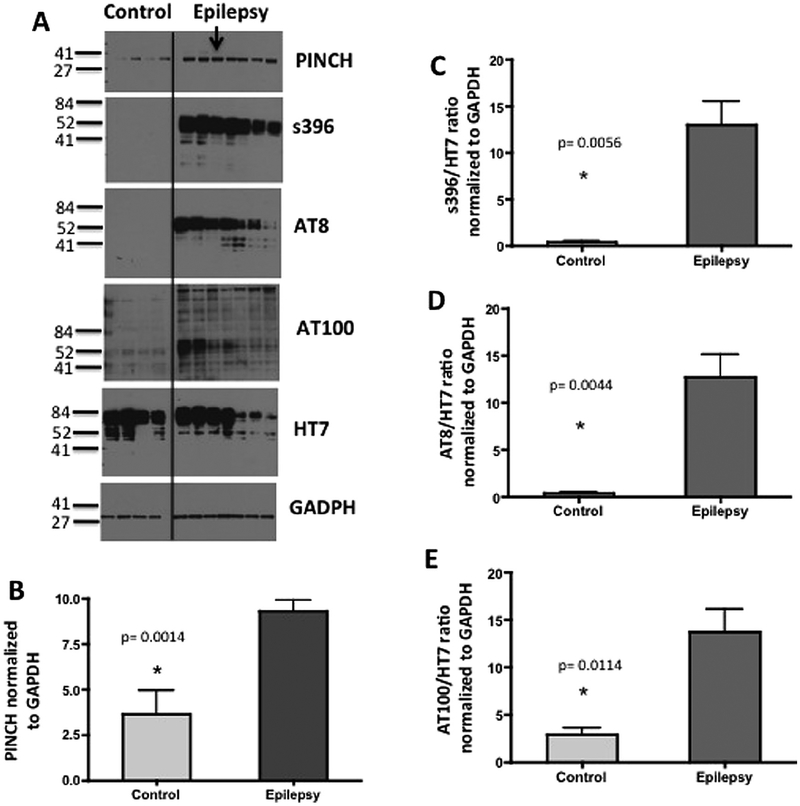
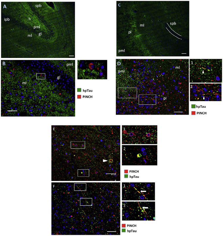
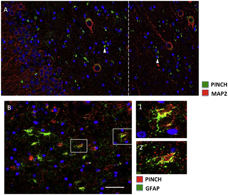
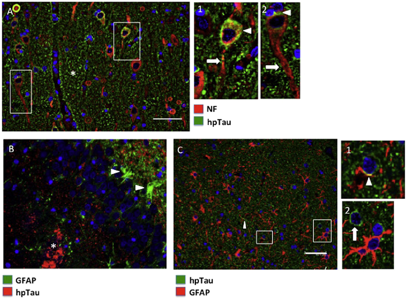
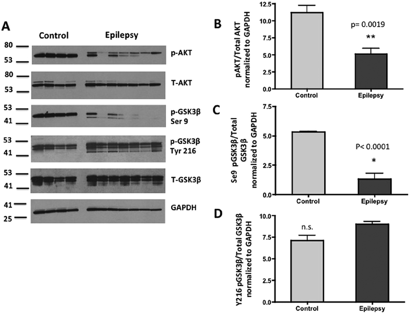
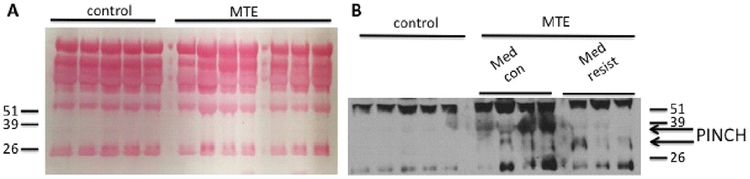
Mounting evidence suggests that inflammation is important in epileptogenesis. Particularly Interesting New Cysteine Histidine-rich (PINCH) protein is a highly conserved, LIM-domain protein known to interact with hyperphosphorylated Tau. We assessed PINCH expression in resected epileptogenic human hippocampi and further explored the relationships among PINCH, hpTau and associated kinases. Resected hippocampal tissue from 7 patients with mesial temporal lobe epilepsy (MTLE) was assessed by Western analyses to measure levels of PINCH and hyperphosphorylated Tau, as well as changes in phosphorylation levels of associated kinases AKT and GSK3β in comparison to normal control tissue. Immunolabeling was also conducted to evaluate PINCH and hpTau patterns of expression, co-localization and cell-type specific expression. Hippocampal PINCH was increased by 2.6 fold in the epilepsy cases over controls and hpTau was increased 10 fold over control. Decreased phospho-AKT and phospho-GSK3β in epilepsy tissue suggested involvement of this pathway in MTLE. PINCH and hpTau co-localized in some neurons in MTLE tissue. While PINCH was expressed by both neurons and astrocytes in MTLE tissue, hpTau was extracellular or associated with neurons. PINCH was absent from the serum of control subjects but readily detectable from the serum of patients with chronic epilepsy. Our study describes the expression of PINCH and points to AKT/GSK3β signaling dysregulation as a possible pathway in hpTau formation in MTLE. In view of the interactions between hpTau and PINCH, understanding the role of PINCH in MTLE may provide increased understanding of mechanisms leading to inflammation and MTLE epileptogenesis and a potential biomarker for drug-resistant epilepsy.

Keywords: Hippocampal sclerosis; Hyperphosphorylated Tau; Mesial temporal lobe epilepsy; PINCH protein.


Figures

Fig. 1.
Fig. 1.
Proposed PINCH signaling pathway involved in hpTau accumulation in MTLE.
Fig. 2.
Fig. 2.
PINCH and hpTau levels are significantly increased in MTLE. (A) Representative Western blot shows increased PINCH and hpTau in epilepsy (n = 7) versus control (n = 4) tissues. Arrow indicates the case with out sclerosis. Membranes containing 20 μg/lane of total protein were probed with anti-PINCH antibody, and anti-Tau antibodies: S396, AT8 and AT100 and HT7 and GAPDH as a loading control. (B) Graphic representation of fold change of PINCH over GAPDH,*p = 0.0014 by unpaired t-test. (C) Graphic representation of fold change of hpTau (S396) over total Tau (HT7),*p = 0.0056 by unpaired t-test. (D) Graphic representation of fold change of hpTau (AT8) over total Tau (HT7), *p = 0.0044 by unpaired t-test. (E) Graphic representation of fold change of hpTau (AT100) over total Tau (HT7), *p = 0.0114 by unpaired t-test.
Fig. 3.
Fig. 3.
Patterns of PINCH and hpTau expression in the hippocampus from representative MTLE cases. (A) hpTau expression pattern (green) in the dentate gyrus. (B) Double immunolabeling for PINCH (red) and hpTau (green) in the dentate gyrus. Inset shows a PINCH immunoreactive cell at the junction of the molecular and granular layers. (C) hpTau pattern (green) in the CA1/2 region of the hippocampus. The white line indicates the suprapyramidal blade of the dentate gyrus. (D) Double immunolabeling of PINCH (red) and hpTau (green) in the CA2 region of the hippocampus. Insets indicate some areas of hpTau/PINCH co-localization (arrowheads) with cellular processes. (E) PINCH (red) and hpTau (green) double immunolabeling in the CA1/2 region of the hippocampus. Arrowhead indicates extracellular accumulation of PINCH and hpTau. Inset 1, PINCH labeling of small neurons. Inset 2, PINCH/hpTau co-localization in a small cell. (F) PINCH and hpTau co-localization in long cellular processes in the CA1/2 region, arrows, insets). Nuclei are labeled blue with DAPI. spb, suprapyramidal blade of dentate gyrus; h, hilus; ipb, infrapyramidal blade of dentate gyrus; pml, polymorphic layer; ml, molecular layer; gl, granular layer; pl, pyramidal layer. Scale bars ≈ 40 μm.
Fig. 4.
Fig. 4.
PINCH expression in neurons and astrocytes in the hippocampus from representative MTLE cases. (A) Double immunolabeling of PINCH (green) and MAP2 (red) in large neurons in the hilus region of dentate gyrus. The white-hatched line indicates the junction of image stitching. PINCH is detected in MAP2 negative smaller cell bodies and processes (arrowheads). (B) PINCH (red) and GFAP (green) co-localize in the CA1/2 region of the hippocampus (insets 1 and 2). Nuclei are labeled blue with DAPI. Scale bars ≈ 40 μm.
Fig. 5.
Fig. 5.
hpTau expression in neurons and astrocytes in the hippocampus from representative MTLE cases. (A) Double immunolabeling of hpTau (green) and neurofilament (red) in the CA1/2 region. Asterisk indicates largely extracellular hpTau. Insets indicate hpTau in neuronal soma (arrowheads) with little to no detection in the axon (arrow). (B) Double immunolabeling of astrocytes (GFAP, green) and hpTau (red). Reactive astrocytes (arrowheads) are adjacent to hpTau, but no co-localization is observed. Asterisk indicates accumulation of hpTau in the polymorphic layer of the dentate gyrus. (C) In the CA1/2 region, GFAP (red) and hpTau (green) co-localization is minimal (inset 1, arrowhead) and two clearly distinct cell populations are labeled (inset 2). Nuclei are labeled blue with DAPI. Scale bars ≈ 40 μm.
Fig. 6.
Fig. 6.
Levels of phosphorylated AKT and GSK3β are significantly decreased in MTLE compared to control. (A) Representative Western blot shows decreased pAKT, pGSK3β-S9 in epilepsy (n = 7) versus control (n = 4) tissues. Arrow indicates the case with out sclerosis. Membranes containing 20 μg/lane of total protein were probed with anti-pAKT serine 473 (ser273), total AKT, pGSK3β serine 9 (ser9), pGSK3β tyrosine 216 (tyr216), total GSK3β antibodies and GAPDH antibody as a loading control. (B) Graphic representation of the ratio of pAKT to total AKT (**p = 0.0019), and pGSK3β-S9 to total GSK3β (p, 0.0001) normalized to GAPDH by unpaired t-test. No significant differences (n.s.) were observed in levels of pGSK3β at tyrosine 216 (Tyr216).
Fig. 7.
Fig. 7.
PINCH is detectable in serum from epilepsy patients. (A) Ponceau red staining of the membrane shows even loading. (B) Western blot shows PINCH levels in epilepsy serum samples (n = 7) versus control (n = 4). Membranes containing 10 μl/lane of IgG/Albumin depleted serum were probed with anti-PINCH antibody.

References

    1. Adiga R, Ozdemir Ahmet Y, Carides Alexandra, et al. Changes in PINCH levels in the CSF of HIV patients correlate with CD4 count and with hpTau. J Neurovirol 2014;20:371–9. - PMC - PubMed
    1. Anthony IC, Bell JE. The Neuropathology of HIV/AIDS. Int Rev Psychiatry 2008;20:15–24. - PubMed
    1. Anthony IC, Ramage SN, Carnie FW, et al. Accelerated Tau deposition in the brains of individuals infected with human immunodeficiency virus-1 before and after the advent of highly active anti-retroviral therapy. Acta Neuropathol 2006;111:529–38. - PubMed
    1. Augustinack JC, Schneider A, Mandelkow EM, et al. Specific tau phosphorylation sites correlate with severity of neuronal cytopathology in Alzheimer’s disease. Acta Neuropathol 2002;103:26–35. - PubMed
    1. Breteler MM, de Groot RR, van Romunde LK, et al. Risk of dementia in patients with Parkinson’s disease, epilepsy, and severe head trauma: a register-based follow-up study. Am J Epidemiol 1995;142:1300–5. - PubMed

MeSH terms

LinkOut - more resources