. 2017 Jan:78:9-24.
doi: 10.1016/j.mcn.2016.11.004. Epub 2016 Nov 10.

Neurexin, Neuroligin and Wishful Thinking coordinate synaptic cytoarchitecture and growth at neuromuscular junctions

Affiliations

Neurexin, Neuroligin and Wishful Thinking coordinate synaptic cytoarchitecture and growth at neuromuscular junctions

Swati Banerjee et al. Mol Cell Neurosci. 2017 Jan.

Abstract

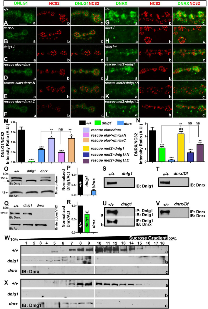
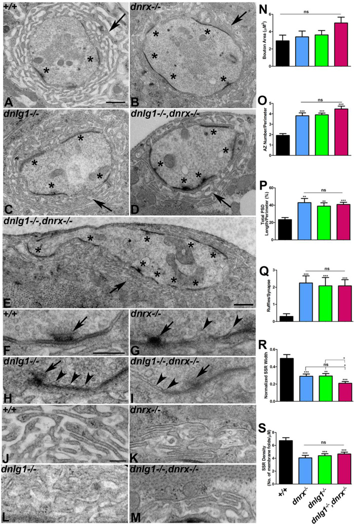
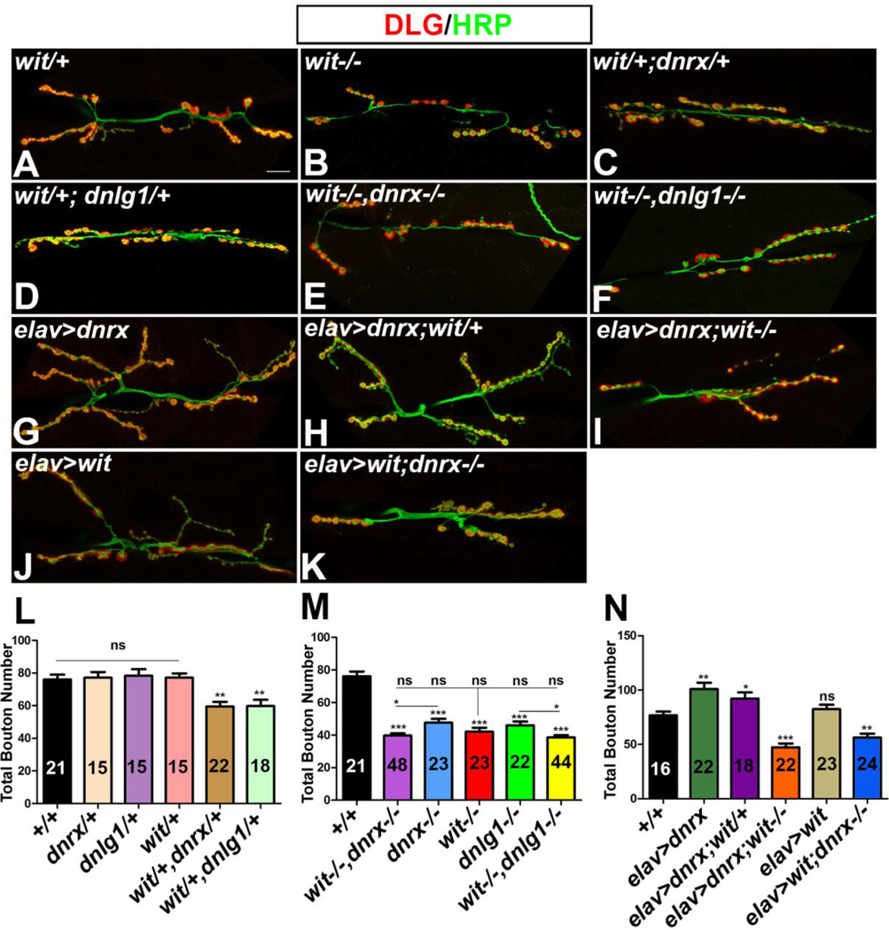
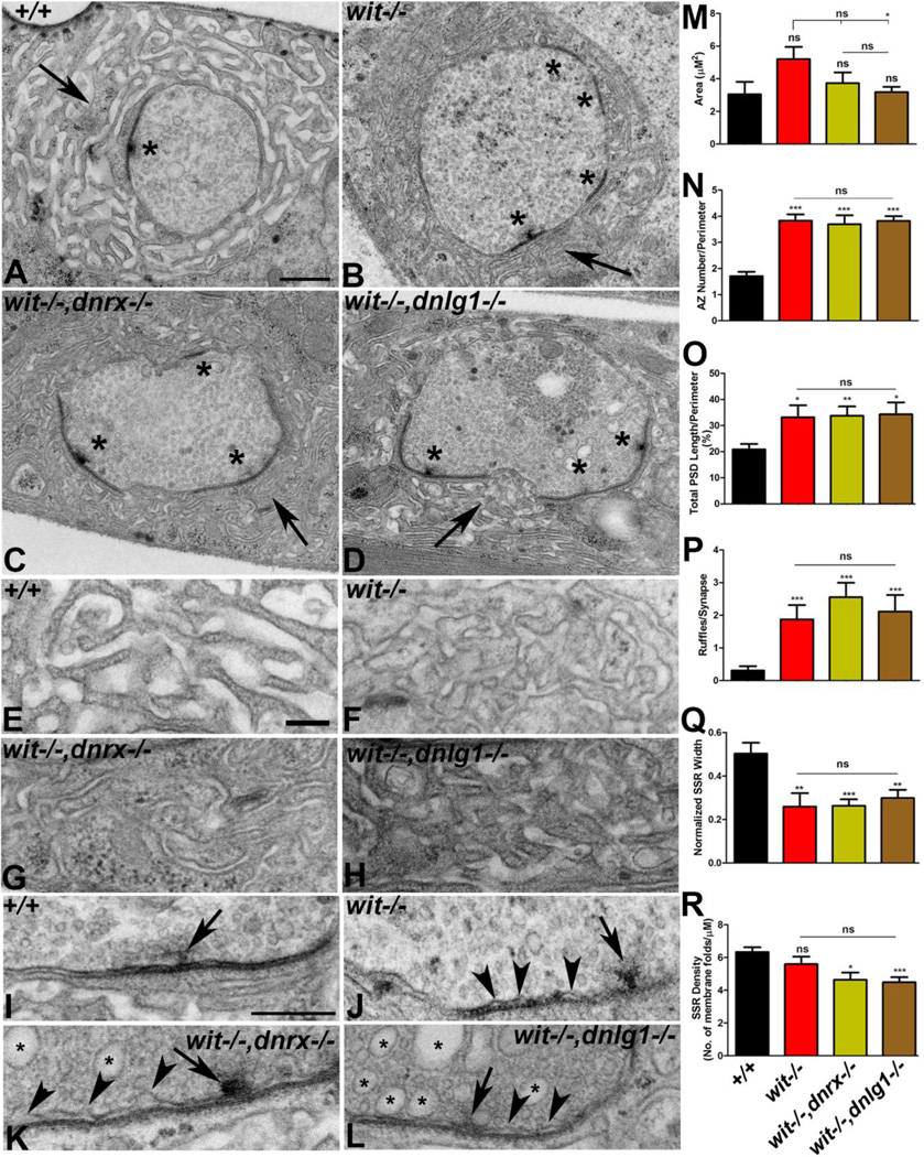
Trans-synaptic interactions involving Neurexins and Neuroligins are thought to promote adhesive interactions for precise alignment of the pre- and postsynaptic compartments and organize synaptic macromolecular complexes across species. In Drosophila, while Neurexin (Dnrx) and Neuroligins (Dnlg) are emerging as central organizing molecules at synapses, very little is known of the spectrum of proteins that might be recruited to the Dnrx/Dnlg trans-synaptic interface for organization and growth of the synapses. Using full length and truncated forms of Dnrx and Dnlg1 together with cell biological analyses and genetic interactions, we report novel functions of Dnrx and Dnlg1 in clustering of pre- and postsynaptic proteins, coordination of synaptic growth and ultrastructural organization. We show that Dnrx and Dnlg1 extracellular and intracellular regions are required for proper synaptic growth and localization of Dnlg1 and Dnrx, respectively. dnrx and dnlg1 single and double mutants display altered subcellular distribution of Discs large (Dlg), which is the homolog of mammalian post-synaptic density protein, PSD95. dnrx and dnlg1 mutants also display ultrastructural defects ranging from abnormal active zones, misformed pre- and post-synaptic areas with underdeveloped subsynaptic reticulum. Interestingly, dnrx and dnlg1 mutants have reduced levels of the Bone Morphogenetic Protein (BMP) receptor Wishful thinking (Wit), and Dnrx and Dnlg1 are required for proper localization and stability of Wit. In addition, the synaptic overgrowth phenotype resulting from the overexpression of Dnrx fails to manifest in wit mutants. Phenotypic analyses of dnrx/wit and dnlg1/wit mutants indicate that Dnrx/Dnlg1/Wit coordinate synaptic growth and architecture at the NMJ. Our findings also demonstrate that loss of Dnrx and Dnlg1 leads to decreased levels of the BMP co-receptor, Thickveins and the downstream effector phosphorylated Mad at the Neuromuscular Junction (NMJ) synapses indicating that Dnrx/Dnlg1 regulate components of the BMP signaling pathway. Together our findings reveal that Dnrx/Dnlg are at the core of a highly orchestrated process that combines adhesive and signaling mechanisms to ensure proper synaptic organization and growth during NMJ development.

Keywords: Neurexin; Neuroligin 1; Neuromuscular junctions; Subsynaptic reticulum; Synaptic growth; Wishful Thinking.


Conflict of interest statement

The authors declare no conflict of interest.

Figures

Figure 1
Figure 1. Interdependence in synaptic clustering of Dnlg1 and Dnrx
(A-L) Confocal micrograhs of 3rd instar larval NMJ labeled with antibodies against Dnlg1 (A-F, green), Dnrx (G-L, green) and active zone protein Bruchpilot (NC82, red, A-L). Scale bar equals 5µM for panels (A-L). (M, N) Quantification of Dnlg1/NC82 (M) and Dnrx/NC82 (N) fluorescence intensity ratio in the specified genotypes. Number of larvae (n) for each genotype under different experimental group was 8. (O) Immunoblots of larval musculature extracts from wild type, dnlg1 and dnrx mutants probed with anti-Dnlg1 (O, arrowhead, 150kDa) showing levels of Dnlg1. Arrow points to a non-specific band in the Dnlg1 immunoblot. Anti-Actin was used as a protein loading control (arrow, 42kDa). (Q) Immunoblots from 3rd instar larval brain lobe/VNC extracts showing levels of Dnrx (Q, arrow, 220kDa) in wild type, dnlg1 and dnrx mutants. (P, R) Quantification of the ratio of band intensities of Dnlg1 (P) and Dnrx (R) with their respective Actin levels used as loading control. (S-V) Adult head lysates of wild type (+/+) and dnlg1 probed with anti-Dnlg1 (S), and wild type (+/+) and dnrx/Df probed with anti-Dnrx (T) antibodies. Head lysates of wild type (+/+) in combination with either dnlg1 or dnrx/Df immunoprecipitated with anti-Dnlg1 (Ua, b) and anti-Dnrx (V). (W, X) 10–45% continuous sucrose density gradient centrifugation of head lysates from the wild type, dnrx and dnlg1 mutants immunoblotted against Dnrx (W) and Dnlg1 (X) show Dnrx and Dnlg1 in overlapping fractions (7–15) in wild type (+/+, compare Wa and Xa). Gradient fractions that correspond up to ~22% sucrose are shown.
Figure 2
Figure 2. Domain-specific requirement of Dnrx in synaptic growth
(A-H) Merged confocal sections from 3rd instar larval NMJ of muscle 6/7 immunostained with antibodies against the presynaptic marker Hrp (green) and the postsynaptic marker Dlg (red) show synaptic growth in wild type (+/+, A), dnrx (B), presynaptic overexpression of Dnrx (C), DnrxΔN (E) and DnrxΔC (G) using elav-Gal4 in the wild type background. Corresponding dnrx mutant rescues using presynaptic overexpression of Dnrx (D), DnrxΔN (F) and DnrxΔC (H). (I) Quantification of total bouton numbers in indicated genotypes. Number of larvae quantified (n) for each genotype is indicated within the bar. Scale bar: 10µM.
Figure 3
Figure 3. Subcellular localization of Dlg is altered in single and double mutants of dnlg1 and dnrx
(A-F) Representative confocal images of NMJ boutons double-stained with anti-Dlg (red) and anti-Hrp (green) antibodies. Dlg localization in wild type (+/+, Aa,b) boutons is concentrated at the SSR. Dlg levels are significantly reduced in dnrx (Ba,b), and display a uniform distribution in dnlg1 (Ca,b) compared to wild type (A). Double mutants of dnlg1,dnrx show reduction in levels of Dlg (Da,b) and a uniform distribution. Dlg localization is rescued when full length Dnlg1 is expressed postsynaptically using mef2-Gal4 (Ea, b). Dlg levels also get restored in dnrx mutants upon overexpression of full length Dnrx presynaptically using elav-Gal4 (Fa,b). (G-I) Quantification of Dlg/Hrp fluorescence intensity ratio in the specified genotypes. Number of larvae (n) for each genotype under different experimental group was 8. Scale bar: 5µM.
Figure 4
Figure 4. Synaptic ultrastructure is altered in dnrx and dnlg1 single and double mutants
(A-D) Ultrastructure of type Ib synaptic boutons in wild type (+/+, A), dnrx (B), dnlg1 (C) and dnlg1,dnrx (D). Note the abundant clear vesicles, electron dense active zones (asterisks), and the SSR localized around the presynaptic bouton (arrow) in wild type control (+/+, A). Ultrastructure of mutant type Ib boutons in dnrx (B), dnlg1 (C) and dnlg1,dnrx (D) frequently display gross morphological defects that include increase in active zone number (asterisks) and length, and reduced thickness and altered SSR morphology (arrow). (E) A giant synaptic bouton in dnrx,dnlg1 that is elongated laterally and have multiple active zones (asterisk) along the perimeter. (F-I) Higher magnification images show morphology of T-bars (arrow) and a tightly apposed pre- and postsynaptic membrane in wild type (+/+, F). A compromised T-bar together with increasingly detached presynaptic membrane with characteristic ruffles (arrowheads) are observed in dnrx (G), dnlg1 (H) and dnrx,dnlg1 (I). (J-M) Higher magnification shows SSR morphology in wild type (+/+, J). Altered SSR morphology that display reduced width, thinner folds, less complexity and convolution is seen in dnrx (K), dnlg1 (L) and dnlg1,dnrx (M). (N-S) Quantification of a variety of pre- and postsynaptic abnormalities including bouton area (N), number of active zone/bouton perimeter (O), total PSD length/bouton perimeter (P), number of membrane ruffles/synapse (Q), normalized SSR width (R) and density of SSR membrane folds/µM (S) are shown in indicated genotypes. Scale bar for (A-E) equals 600nm and (F-M) equals 200nm. Number of synaptic boutons (n) from three larvae of each genotype were quantified for morphometric analyses: In wild type (n=15), dnrx (n=16), dnlg1 (n=21) and dnlg1,dnrx (n=17).
Figure 5
Figure 5. Wit localization and stability is dependent on Dnrx and Dnlg1
(A-D) Immunoblots of larval musculature extracts from wild type (+/+), dnlg1, dnrx and wit mutants probed with anti-Wit antibodies (100kDa, A) showing levels of Wit. Anti-Actin was used as a protein loading control. Quantification of the ratio of band intensities of Wit with respect to Actin levels as shown in specified genotypes (B). qRTPCR analysis of wit mRNA levels in indicated genotypes (C). Sucrose density gradient centrifugation of head lysates from the wild type, dnrx and dnlg1 mutants immunoblotted against Wit (D) show Wit distribution in fractions (4–15) in wild type (+/+, Da). A decrease in Wit levels and shift in its distribution is observed in dnlg1 (Db) and dnrx/Df (Dc) gradient fractions. (E, F) 3rd instar larval NMJ of wild type (+/+, E) and wit (F) show localization of Dnrx (Ea,c and Fa,c) and NC82 (Eb,c and Fb,c). (G, H) 3rd instar larval NMJ of wild type (+/+, G) and wit (H) co-stained with anti-Dnlg1 (Ga,c and Ha,c) and anti-BRP (NC82, Eb,c and Fb,c). (I-L) NMJ of elav-Gal4; UAS-wit (I, K) show Wit localization (Ib,c and Kb,c) with respect to Dnrx (Ia,c) and Dnlg1 (Ka,c). NMJ of elav-Gal4; UAS-wit;dnrx−/− (J) and elav-Gal4;UASwit;dnlg1−/− (L) show significant reduction in Wit levels (Jb,c and Lb,c) both in dnrx and dnlg1 mutant backgrounds, respectively. Scale bar equals 5µM (E-L).
Figure 6
Figure 6. Genetic interactions between wit,dnrx and wit,dnlg1
(A-K) Confocal micrograph of 3rd instar larval NMJ labeled with anti-Hrp (green) and anti-Dlg (red) show reduced synaptic growth in single mutants of wit (B), trans-heterozygotes of wit,dnrx (C) and wit,dnlg1 (D), and double mutants of wit,dnrx (E) and wit,dnlg1 (F) compared to wit/+ heterozygotes (A). (G-K) Overgrowth of NMJ boutons caused by presynaptic overexpression of Dnrx (G) is not suppressed by loss of one copy of wit (H). Overgrowth phenotype resulting from presynaptic Dnrx overexpression is attenuated by loss of both copies of wit (I) and resembles the wit mutant phenotype (B). Presynaptic overexpression of Wit does not cause overgrowth of NMJ boutons (J) and fails to rescue the synaptic undergrowth phenotype of dnrx (K). (L-N) Quantification of the NMJ bouton number at A3 in muscles 6 and 7 of designated genotypes. Number of larvae quantified (n) is indicated within the bars representing various genotypic combinations. Scale bar equals 10µM.
Figure 7
Figure 7. Pre- and postsynaptic abnormalities resulting from individual loss of wit and it’s combined loss with dnrx and dnlg1
(A-D) Low magnification TEM images of wild type (+/+, A), wit (B), wit,dnrx (C) and wit,dnlg1 (D). Ultrastructure of mutant boutons (B-D) show severely altered SSR morphology (arrows), decreased SSR width, increased PSD length, and increased number of active zones (asterisks) compared to wild type (+/+, A). (E-H) Higher magnification ultrastructural images highlighting the SSR morphology of wild type (+/+, E), wit (F), wit,dnrx (G) and wit,dnlg1 (H). (I-L) Electron micrograph showing pre- and postsynaptic membrane and T-bar (arrow) morphology in wild type (+/+, I), wit (J), wit,dnrx (K) and wit,dnlg1 (L). Note the presence of presynaptic membrane ruffles in mutants (arrowheads, J-L) and large synaptic vesicles (arrows) in double mutants of wit,dnrx (K) and wit,dnlg1 (L). (M-R) Quantification of bouton area (M), number of active zone/perimeter (N), total PSD length/perimeter (O), number of membrane ruffles/synapse (P), normalized SSR width (Q) and density of SSR membrane folds/µM (R) as shown in specified genotypes. Scale bar for (A-D) equals 600nm and (E-L) equals 200nm.
Figure 8
Figure 8. Thick Veins and pMad levels are reduced at dnrx and dnlg1 mutant NMJ synapses
(A-D) Confocal images of NMJ boutons co-labeled with anti-Tkv (red) and anti-Hrp (green) antibodies. Tkv localization in wild type boutons (+/+, Aa,b), dnrx/Df (Ba,b), dnlg1 (Ca,b) and wit (Da,b).. (E-H) Confocal images of NMJ boutons co-labeled with anti-pMad (red) and anti-Hrp (green) in wild type (+/+, Ea,b), dnrx/Df (Fa,b), dnlg1 (Ga,b) and wit (Ha,b).. (I-N) Confocal micrographs of 3rd instar larval NMJ labeled with anti-Hrp (green) and anti-Dlg (red) showing synaptic growth in Mad/+ (I), double-heterozygotes of Mad;dnrx (J), Mad;dnlg1 (K), Mad−/− (L), and double mutants of Mad;dnrx (M) and Mad;dnlg1 (N). (O-Q) Quantification of Tkv/Hrp (O) and pMad/Hrp (P) fluorescence intensity ratio in the specified genotypes. Number of larvae (n) analyzed for each genotype was 8. Number of synaptic boutons (Q) in stated genotypes. Number of larvae quantified (n) is indicated within the bars representing various genotypic combinations. (R) A model depicting the interactions between Dnrx and Dnlg1 with various components of the BMP signaling pathway at the trans-synaptic interface of NMJ. Optimal BMP signaling requires proper trans-synaptic adhesion and stability of various BMP receptors to guarantee coordinated synaptic growth at the NMJ during development.

References

    1. Aberle H, Haghighi AP, Fetter RD, McCabe BD, Magalhaes TR, Goodman CS. wishful thinking encodes a BMP type II receptor that regulates synaptic growth in Drosophila. Neuron. 2002;33:545–558. - PubMed
    1. Aoto J, Foldy C, Ilcus SM, Tabuchi K, Sudhof TC. Distinct cirucuit-dependent functions of presynaptic neurexin-3 at GABAergic and glutamatergic synapses. Nat Neuroscience. 2015;18:997–1007. - PMC - PubMed
    1. Ashley J, Packard M, Ataman B, Budnik V. Fasciclin II signals new synapse formation through amyloid precursor protein and scaffolding protein dX11/Mint. J Neurosci. 2005;25:5943–5955. - PMC - PubMed
    1. Ball RW, Warren-Paquin M, Tsurudome K, Liao EH, Elazzouzi F, Cavanagh C, An BS, Wang TT, White JH, Haghighi AP. Retrograde BMP signaling controls synaptic growth at the NMJ by regulating trio expression in motor neurons. Neuron. 2010;66:536–549. - PubMed
    1. Banerjee S, Riordan M, Bhat MA. Genetic aspects of autism spectrum disorders: insights from animal models. Frontiers in cellular neuroscience. 2014;8:58. - PMC - PubMed

MeSH terms