. 2016 Dec:48:51-62.
doi: 10.1016/j.dnarep.2016.10.011. Epub 2016 Oct 31.

Identification of SUMO modification sites in the base excision repair protein, Ntg1

Affiliations

Identification of SUMO modification sites in the base excision repair protein, Ntg1

Daniel B Swartzlander et al. DNA Repair (Amst). 2016 Dec.

Abstract

DNA damaging agents are a constant threat to genomes in both the nucleus and the mitochondria. To combat this threat, a suite of DNA repair pathways cooperate to repair numerous types of DNA damage. If left unrepaired, these damages can result in the accumulation of mutations which can lead to deleterious consequences including cancer and neurodegenerative disorders. The base excision repair (BER) pathway is highly conserved from bacteria to humans and is primarily responsible for the removal and subsequent repair of toxic and mutagenic oxidative DNA lesions. Although the biochemical steps that occur in the BER pathway have been well defined, little is known about how the BER machinery is regulated. The budding yeast, Saccharomyces cerevisiae is a powerful model system to biochemically and genetically dissect BER. BER is initiated by DNA N-glycosylases, such as S. cerevisiae Ntg1. Previous work demonstrates that Ntg1 is post-translationally modified by SUMO in response to oxidative DNA damage suggesting that this modification could modulate the function of Ntg1. In this study, we mapped the specific sites of SUMO modification within Ntg1 and identified the enzymes responsible for sumoylating/desumoylating Ntg1. Using a non-sumoylatable version of Ntg1, ntg1ΔSUMO, we performed an initial assessment of the functional impact of Ntg1 SUMO modification in the cellular response to DNA damage. Finally, we demonstrate that, similar to Ntg1, the human homologue of Ntg1, NTHL1, can also be SUMO-modified in response to oxidative stress. Our results suggest that SUMO modification of BER proteins could be a conserved mechanism to coordinate cellular responses to DNA damage.

Keywords: Base excision repair (BER); NTHL1; Ntg1; SUMO; Sumoylation.


Conflict of interest statement

The authors declare that there are no conflicts of interest.

Figures
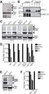
Figure 1
Figure 1. Sumoylation of Ntg1 is conserved and mediated by Siz1/2
A. Wildtype S. cerevisiae cells expressing Ntg1-TAP were exposed to 0 (−) or 20 mM (+) H2O2 for 1 hour at 30°C. Cells were pelleted, lysed, and immunoblotted to detect TAP-tagged Ntg1. Bands corresponding to post-translationally modified Ntg1 including SUMO-modified Ntg1 (24) are indicated by Ntg1-TAP*. B. Colon adenocarcinoma cells (HT29) were transfected with NTHL1-Flag or empty Flag vector and treated with 0 (−) or 125 µM (+) H2O2 for 15 minutes at 37°C. Cells were lysed, immunoprecipitated with Flag antibodies and both the Input and Flag IP fractions were subjected to immunoblotting. An IgG bead alone immunoprecipitation was included as a control. The blot was probed with NTHL1 and SUMO-2/3 antibodies as indicated. C. Wildtype (WT), siz1Δ, siz2Δ, siz1Δsiz2Δ, ulp1-ts, or ulp2Δ cells were transformed with a plasmid expressing Ntg1-TAP. Cells were (C) exposed to 20 mM hydrogen peroxide or (E) not treated. Cells were incubated at 30°C except ulp1-ts cells which were shifted to the non-permissive temperature of 37°C. Each sample was lysed, immunoblotted, and bands were quantified. Nonadjacent lanes in the same image are separated by a black line. D. The data from (C) were quantitated. The total amount of Ntg1-TAP including unmodified and modified Ntg1-TAP was set to 100% (Ntg1) and the fraction of signal present in bands (Total Ntg1 Signal %) corresponding to the size consistent with Mono-, Di-, and Tri-sumoylation is plotted on a log scale. Results shown are the average of two independent experiments. Error bars represent SEM. E. To examine sumoylation of Ntg1 in the absence of oxidative damage, ulp1-ts and ulp2Δ cells expressing Ntg1-TAP were analyzed to detect any modified Ntg1 species (Ntg1-TAP*). F. The data from (E) were quantitated. The total amount of Ntg1-TAP including unmodified and modified Ntg1-TAP was set to 100% (Ntg1) and the fraction of signal present in bands (Total Ntg1 Signal %) corresponding to the size consistent with Mono-, Di-, and Tri-sumoylation is plotted on a log scale. Results shown are the average of two independent experiments. Error bars represent SEM.
Figure 2
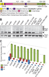
Figure 2. Identification of sumoylation sites in Ntg1
A. A domain schematic of Ntg1 is shown with the following functional motifs/domains indicated: The Mitochondrial Targeting Sequence (MTS) in yellow, the classical Nuclear Localization Signal (cNLS) in dark blue, the Catalytic Domain in purple. The Catalytic Lysine, K243, is depicted as a black bar. The central region of Ntg1 that is homologous to E. coli Endonuclease III is shown in tan (amino acids 95–335) while the non-conserved N- and C-terminal domains are indicted in magenta (amino acids 1–94) and green (amino acids 336–399), respectively. Putative Consensus Sumoylation Sites are shown as red bars and Putative Non-Consensus Sumoylation Sites are shown as grey bars. B. A series of Ntg1 variants with candidate SUMO modification sites altered from lysine to arginine were generated and expressed in temperature sensitive ulp1 cells. Cells were treated with 20 mM hydrogen peroxide for 1 hour at 30°C, lysed, and immunoblotted to detected Ntg1-TAP and modified Ntg1-TAP (Ntg1-TAP*). Nonadjacent lanes in the same image are separated by white space. C. Results from (B) were quantitated. For each Ntg1 variant, the percent of total Ntg1-TAP signal present in the band corresponding to the size of Mono-, Di-, and Tri-sumoylation (indicated as Percent Sumoylation) is plotted on a log scale.
Figure 3
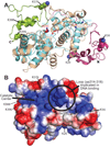
Figure 3. Homlogy model of Ntg1
A. A homology model of Ntg1 shown as a ribbon diagram was generated as described in Materials and Methods. The model is overlaid on the E. coli Ntg1 homologue, Endonuclease III, structure (cyan, PDB ID: 2ABK). The Ntg1 catalytic domain (amino acids 95–335; tan), N-terminal domain (amino acids 1–94; magenta), C-terminal domain (amino acids 336–399; green), catalytic amino acid of Ntg1 (K243, red) and Endonuclease III (K120, blue) are shown in addition to Endonuclease III amino acids D138, important for catalysis, and K191, implicated in DNA binding (69), and the corresponding amino acids in Ntg1 (D262 and N318, respectively). The five consensus sumoylation sites (K20, K38, K376, K388, and K396) are shown as balls and indicated by the labeling. B. An electrostatic model of Ntg1 is shown based on the homology model. Positive and negative residues are colored in blue and red, respectively. White indicates neutral residues. The loop containing residues 314–318, indicated by a circle, has been implicated in DNA binding by Endo III (69). The catalytic center is indicated by a bold black line and the five consensus sumoylation sites are labeled and indicated by black lines. Residues 20, 376, 388, and 396 are located on the back face of the model and are indicated by black dotted lines.
Figure 4
Figure 4. Functional analysis of Ntg1 variant
A. A schematic of the substrate employed for the in vitro cleavage assay, which contains dihydrouracil (DHU) embedded in a 31mer oligo, illustrating the substrate and expected products of the cleavage reaction is shown. B. Recombinant E. coli Endonuclease III (Endo III), and Ntg1 variants, His6-Ntg1, His6-ntg1(K->R)5, catalytically inactive ntg1 (His6-ntg1Δcat), were employed for the in vitro cleavage assay. Increasing amounts of recombinant protein (5–50 ng) were added to radioactively-labeled substrate. Oligonucleotide Cleavage Products were electrophoresed and subjected to phosphorimager analysis. The Control lane shows the substrate with no added protein. The positive control is addition of 50 ng of E. coli Endo III. The position of the labeled product generated by cleavage (Cleavage Product) is indicated. Random degradation product is indicated by an asterisk (*). Nonadjacent lanes in the same image are separated by black lines. Results shown in (B) are representative of three independent experiments. C. Quantification of Cleavage Product generated for each Ntg1 variant from three independent experiments. Results are shown as Percent DHU Cleaved. Error bars represent standard deviation in the data.
Figure 5
Figure 5. Functional analysis of ntg1ΔSUMO in DNA damage pathways
A. Wildtype cells expressing Ntg1-TAP were exposed to hydrogen peroxide (H2O2), methyl methanesulfonate (MMS), or were not treated (NT) and lysed. Lysate was subjected to immunoblotting to detect Ntg1-TAP and modified forms of Ntg1-TAP (Ntg1-TAP*). B. Cells with either a full complement of wildtype (WT) DNA repair pathways or deficient in both base excision repair and nucleotide excision repair (B-/N-) were employed. As described in Materials and Methods, the genotype for B-/N- cells (DSC0369) is ntg1Δntg2Δapn1Δrad1Δ. Both the WT and B-/N- cells were engineered to express ntg1ΔSUMO and compared to cells expressing NTG1 or lacking Ntg1 (ntg1Δ). Cultures were 5-fold serially diluted and spotted onto rich media or rich media containing 0.005% MMS and incubated at 30°C for 4 days. Pictures were taken at Day 2 and Day 4. C/D. The same samples as shown in (B) with either intact DNA repair pathways (WT) (blue diamond) or deficient in base excision repair and nucleotide excision repair (B-/N-), denoted by an *, contain wildtype NTG1 (red square), or ntg1ΔSUMO (green triangle), or lack Ntg1 (ntg1Δ) (purple X) at the endogenous NTG1 locus. The genotype for B-/N- is ntg1Δntg2Δapn1Δrad1Δ (DSC0369). Cells were grown in liquid culture with No MMS (C) or with 0.01% MMS (D) for 48 hours. OD600 readings were taken every 30 minutes and plotted vs time. Results shown in (B, C, and D) are representative of at least three independent experiments.

Similar articles

Cited by

References

    1. Altieri F, Grillo C, Maceroni M, Chichiarelli S. DNA damage and repair: from molecular mechanisms to health implications. Antioxid Redox Signal. 2008;10(5):891–937. - PubMed
    1. Fortini P, Pascucci B, Parlanti E, D'Errico M, Simonelli V, Dogliotti E. The base excision repair: mechanisms and its relevance for cancer susceptibility. Biochimie. 2003;85(11):1053–1071. - PubMed
    1. Boesch P, Weber-Lotfi F, Ibrahim N, Tarasenko V, Cosset A, Paulus F, et al. DNA repair in organelles: Pathways, organization, regulation, relevance in disease and aging. Biochim Biophys Acta. 2011;1813(1):186–200. - PubMed
    1. Kryston TB, Georgiev AB, Pissis P, Georgakilas AG. Role of oxidative stress and DNA damage in human carcinogenesis. Mutation research. 2011;711(1–2):193–201. - PubMed
    1. Pan Y. Mitochondria, reactive oxygen species, and chronological aging: a message from yeast. Experimental gerontology. 2011;46(11):847–852. - PubMed

Web References

    1. http://www.abgent.com/sumoplot Date Last used: 8/4/16.

MeSH terms

LinkOut - more resources