

Selective JAK3 Inhibitors with a Covalent Reversible Binding Mode Targeting a New Induced Fit Binding Pocket
- PMID: 27840070
- PMCID: PMC5119931
- DOI: 10.1016/j.chembiol.2016.10.008
Selective JAK3 Inhibitors with a Covalent Reversible Binding Mode Targeting a New Induced Fit Binding Pocket
Abstract
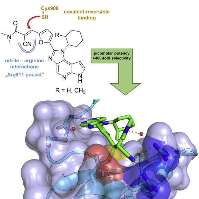
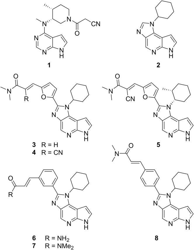
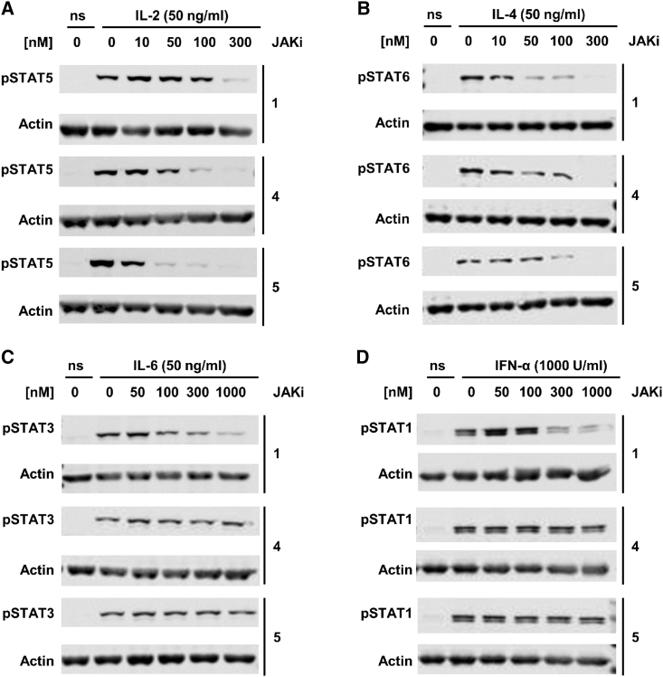
Janus kinases (JAKs) are a family of cytoplasmatic tyrosine kinases that are attractive targets for the development of anti-inflammatory drugs given their roles in cytokine signaling. One question regarding JAKs and their inhibitors that remains under intensive debate is whether JAK inhibitors should be isoform selective. Since JAK3 functions are restricted to immune cells, an isoform-selective inhibitor for JAK3 could be especially valuable to achieve clinically more useful and precise effects. However, the high degree of structural conservation makes isoform-selective targeting a challenging task. Here, we present picomolar inhibitors with unprecedented kinome-wide selectivity for JAK3. Selectivity was achieved by concurrent covalent reversible targeting of a JAK3-specific cysteine residue and a ligand-induced binding pocket. We confirmed that in vitro activity and selectivity translate well into the cellular environment and suggest that our inhibitors are powerful tools to elucidate JAK3-specific functions.
Keywords: JAK3; Janus kinases; chemical probe; covalent reversible inhibitor; kinome selectivity.
Copyright © 2016 The Authors. Published by Elsevier Ltd.. All rights reserved.
Figures
References
-
- Ahearn S.P., Christopher M., Jung J., Pu Q., Rivkin A., Scott M.E., Witter D.J., Woo H.C., Cash B., Dinsmore C. 2013. Pyrrolopyrimidines as Janus Kinase Inhibitors. WO Patent 2013/085802.https://www.google.com/patents/WO2013085802A1?cl=enIt
-
- Bauer S.M., Gehringer M., Laufer S.A. A direct enzyme-linked immunosorbent assay (ELISA) for the quantitative evaluation of Janus Kinase 3 (JAK3) inhibitors. Anal. Methods. 2014;6:8817–8822.
-
- Chrencik J.E., Patny A., Leung I.K., Korniski B., Emmons T.L., Hall T., Weinberg R.A., Gormley J.A., Williams J.M., Day J.E. Structural and thermodynamic characterization of the TYK2 and JAK3 kinase domains in complex with CP-690550 and CMP-6. J. Mol. Biol. 2010;400:413–433. - PubMed
-
- Flanagan M.E., Blumenkopf T.A., Brissette W.H., Brown M.F., Casavant J.M., Poa C.S., Doty J.L., Elliott E.A., Fisher M.B., Hines M. Discovery of CP-690'550: a potent and selective Janus Kinase (JAK) inhibitor for the treatment of autoimmune diseases and organ transplant rejection. J. Med. Chem. 2010;53:8468–8484. - PubMed
-
- Gehringer M., Pfaffenrot E., Bauer S., Laufer S.A. Design and synthesis of tricyclic JAK3 inhibitors with picomolar affinities as novel molecular probes. ChemMedChem. 2014;9:277–281. - PubMed
MeSH terms
Substances
LinkOut - more resources
Full Text Sources
Other Literature Sources
Chemical Information
