Cystic pancreatic lesions: From increased diagnosis rate to new dilemmas
- PMID: 27840080
- PMCID: PMC5546617
- DOI: 10.1016/j.diii.2016.08.017
Cystic pancreatic lesions: From increased diagnosis rate to new dilemmas
Abstract
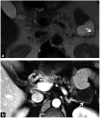
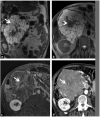
Cystic pancreatic lesions vary from benign to malignant entities and are increasingly detected on cross-sectional imaging. Knowledge of the imaging appearances of cystic pancreatic lesions may help radiologists in their diagnostic reporting and management. In this review, we discuss the morphologic classification of these lesions based on a diagnostic algorithm as well as the management of these lesions.
Keywords: Cyst; IPMN; MRI; Pancreas.
Copyright © 2016 Editions françaises de radiologie. Published by Elsevier Masson SAS. All rights reserved.
Figures


References
-
- Heyn C, Sue-Chue-Lam D, Jhaveri K, Haider MA. MRI of the pancreas: problem solving tool. J Magn Reson Imaging. 2012;36:1037–51. - PubMed
-
- Lewandrowski K, Warshaw A, Compton C. Macrocystic serous cystadenoma of the pancreas: a morphologic variant differing from microcystic adenoma. Hum Pathol. 1992;23:871–5. - PubMed
-
- Zhang XM, Mitchell DG, Dohke M, Holland GA, Parker L. Pancreatic cysts: depiction on single-shot fast spin-echo MR images. Radiology. 2002;223:547–53. - PubMed
-
- Kopelman Y, Groissman G, Fireman Z. Cystic lesion of the pancreas. Gastrointest Endosc. 2007;65:1074–5. - PubMed
Publication types
MeSH terms
Grants and funding
LinkOut - more resources
Full Text Sources
Other Literature Sources
Medical

