. 2016 Dec;14(6):4999-5006.
doi: 10.3892/mmr.2016.5888. Epub 2016 Oct 25.

Identification of HLA‑A*1101‑restricted cytotoxic T lymphocyte epitopes derived from epidermal growth factor pathway substrate number 8

Affiliations

Identification of HLA‑A*1101‑restricted cytotoxic T lymphocyte epitopes derived from epidermal growth factor pathway substrate number 8

Huifang Lu et al. Mol Med Rep. 2016 Dec.

Abstract

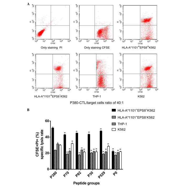
Epidermal growth factor receptor pathway substrate 8 (EPS8) is critical in the proliferation, progression and metastasis of solid and hematological types of cancer, and thus constitutes an ideal target for cancer immunotherapy. The present study aimed to identify human leukocyte antigen (HLA)‑A*1101‑restricted cytotoxic T lymphocyte (CTL) epitopes from EPS8 and characterize their immunotherapeutic efficacy in vitro. Two computer‑based algorithms were used to predict native EPS8 epitopes with potential high binding affinity to the HLA‑A*1101 molecule, which is the HLA‑A allele with the highest frequency in the Chinese population. The peptide‑induced cytokine production from the CTLs was examined using enzyme‑linked immunosorbent spot analysis. The cytotoxic effects on cancer cells by CTLs primed with the identified peptides were examined using flow cytometry. A total of five peptides, designated as P380, P70, P82, P30 and P529, presented with high affinity towards the HLA‑A*1101 molecule. In response to stimulation by these five peptides, enhanced secretion of interferon‑γ from the CTLs and increased cytolytic capabilities of the CTLs toward cancer cells were noted, with the most potent effects observed from the P380 peptide. Taken together, the present study identified five potential CTL epitopes from EPS8. Among these, P380 presented with the highest therapeutic efficacy in vitro. These peptides may benefit the development of EPS8‑based immunotherapy for the treatment of HLA‑A*1101‑positive hematological malignancies.


Figures

Figure 1.
Figure 1.
Phenotypic characterization of the expression of EPS8 and human leukocyte antigen-A*1101 in different target cells. The expression levels of EPS8 in the indicated target cells were detected using western blot analysis. GAPDH was detected as the loading control. (A) Representative western blot. (B) Quantification of relative expression levels of EPS8. EPS8, epidermal growth factor receptor pathway substrate 8.
Figure 2.
Figure 2.
Detection of the cell-surface expression of HLA-A*1101 in indicated target cells using flow cytometry. The blue histogram represents staining with PE-conjugated isotype-matched IgG; the red histogram represents staining with PE-conjugated HLA-ABC. (A) Representative flow cytometry data showed that K562 and THP-1 were negative for the expression of HLA-A*1101. (B) Representative flow cytometry data showed that HLA-A*1101+EPS8+ K562 and HLA-A*1101+EPS8 K562 were positive for the expression of HLA-A*1101. EPS8, epidermal growth factor receptor pathway substrate 8; HLA, human leukocyte antigen; PE, phycoerythrin.
Figure 3.
Figure 3.
Knocking down the expression of EPS8 in HLA-A*110+ K562 cells by siRNA transient transfection. The HLA-A*1101+ K562 cells were transiently transfected with either control siRNA or si-h-EPS8 101–103) At (A) 24, (B) 48 and (C) 72 h post-transfection, the expression levels of EPS8 in the cells were examined using western blot analysis and quantified, as shown in graphs for the (D) 24, (E) 48 and (F) 72 h groups. β-actin was detected as a loading control. *P<0.05 and **P<0.01, compared with the NC group. The transfection effect at 48 h was more marked. EPS8, epidermal growth factor receptor pathway substrate 8; HLA, human leukocyte antigen; siRNA, small interfering RNA; si-h-EPS8 101–103; EPS8-targeting siRNA 101–103; NC, negative control.
Figure 4.
Figure 4.
Production of IFN-γ by PBMCs primed with different peptides. Peptide-primed PBMCs were exposed to irradiated EPS8-presenting HLA-A*1101+ K562 cells and the production of IFN-γ was detected using an ELISPOT assay. PHA treatment was used as a positive control; P0-CTLs or P-CTLs were used as negative controls. (A) Representative ELISPOT images from each condition is shown. (B) Quantification on the numbers of spot-forming cells is shown. *P<0.05 and **P<0.001, compared with the P380 group. IFN-γ interferon-γ; PBMCs, peripheral blood mononuclear cells; EPS8, epidermal growth factor receptor pathway substrate 8; HLA, human leukocyte antigen; P0, PBMCs with no peptide priming; P-CTL, PBMCs not exposed to EPS8-presenting HLA-A*1101+ K562 cells; ELISPOT, enzyme-linked immunospot; X–VIVO, X–VIVO15 serum-free medium; CTL, control; P, P380.
Figure 5.
Figure 5.
Cytotoxicity of PBMCs primed with different peptides. Peptide-primed PBMCs (effector cells) were mixed with the indicated CFSE-labeled target cells at an effector to target ratio of 40:1. The cell mixture was stained with PI, and CFSE+PI+ dead target cells were detected using flow cytometry. (A) Representative flow images of cytolysis by the P380-primed PBMCs. (B) Quantification of the percentage of CFSE+PI+ dead cells in the peptide-primed PBMCs on the indicated target cells. **P<0.001, compared with the P380 group. PBMCs, peripheral blood mononuclear cells; EPS8, epidermal growth factor receptor pathway substrate 8; HLA, human leukocyte antigen; CTL, cytotoxic T cell; CFSE, carboxyfluorescein succinimidyl ester; PI, propidium iodide.

References

    1. Vardiman JW, Thiele J, Arber DA, Brunning RD, Borowitz MJ, Porwit A, Harris NL, Le Beau MM, Hellström-Lindberg E, Tefferi A, Bloomfield CD. The 2008 revision of the World Health Organization (WHO) classification of myeloid neoplasms and acute leukemia: Rationale and important changes. Blood. 2009;114:937–951. doi: 10.1182/blood-2009-03-209262. - DOI - PubMed
    1. Fielding AK, Richards SM, Chopra R, Lazarus HM, Litzow MR, Buck G, Durrant IJ, Luger SM, Marks DI, Franklin IM, et al. Outcome of 609 adults after relapse of acute lymphoblastic leukemia (ALL); an MRC UKALL12/ECOG 2993 study. Blood. 2007;109:944–950. doi: 10.1182/blood-2006-05-018192. - DOI - PubMed
    1. Schmid C, Labopin M, Nagler A, Niederwieser D, Castagna L, Tabrizi R, Stadler M, Kuball J, Cornelissen J, Vorlicek J, et al. Treatment, risk factors, and outcome of adults with relapsed AML after reduced intensity conditioning for allogeneic stem cell transplantation. Blood. 2012;119:1599–1606. doi: 10.1182/blood-2011-08-375840. - DOI - PubMed
    1. Bocchia M, Defina M, Aprile L, Sicuranza A. Peptide vaccines for hematological malignancies: A missed promise? Int J Hematol. 2014;99:107–116. doi: 10.1007/s12185-013-1497-3. - DOI - PubMed
    1. Mohamed YS, Dunnion D, Teobald I, Walewska R, Browning MJ. In vitro evaluation of human hybrid cell lines generated by fusion of B-lymphoblastoid cells and ex vivo tumour cells as candidate vaccines for haematological malignancies. Vaccine. 2012;30:6578–6587. doi: 10.1016/j.vaccine.2012.08.032. - DOI - PubMed

MeSH terms