. 2016 Nov 14:6:36930.
doi: 10.1038/srep36930.

Glutathione-conjugating and membrane-remodeling activity of GDAP1 relies on amphipathic C-terminal domain

Affiliations

Glutathione-conjugating and membrane-remodeling activity of GDAP1 relies on amphipathic C-terminal domain

Nina Huber et al. Sci Rep. .

Abstract

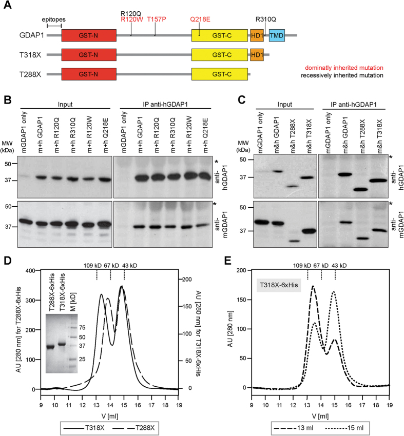
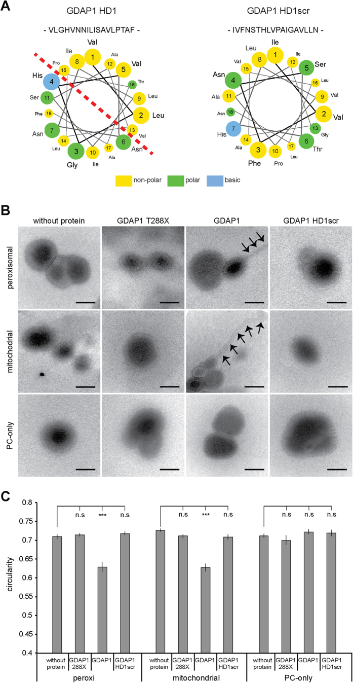
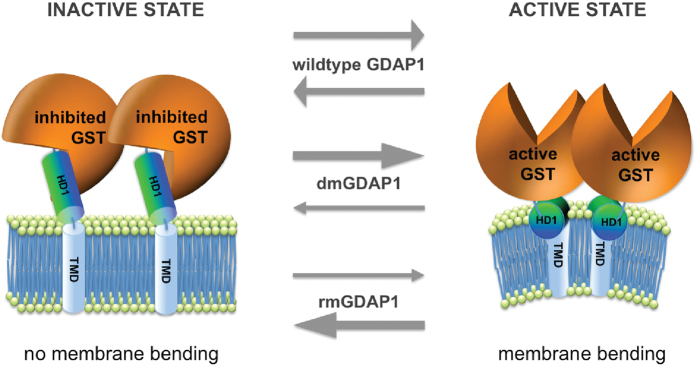
Mutations in the ganglioside-induced differentiation associated protein 1 (GDAP1) cause severe peripheral motor and sensory neuropathies called Charcot-Marie-Tooth disease. GDAP1 expression induces fission of mitochondria and peroxisomes by a currently elusive mechanism, while disease causing mutations in GDAP1 impede the protein's role in mitochondrial dynamics. In silico analysis reveals sequence similarities of GDAP1 to glutathione S-transferases (GSTs). However, a proof of GST activity and its possible impact on membrane dynamics are lacking to date. Using recombinant protein, we demonstrate for the first time theta-class-like GST activity for GDAP1, and it's activity being regulated by the C-terminal hydrophobic domain 1 (HD1) of GDAP1 in an autoinhibitory manner. Moreover, we show that the HD1 amphipathic pattern is required to induce membrane dynamics by GDAP1. As both, fission and GST activities of GDAP1, are critically dependent on HD1, we propose that GDAP1 undergoes a molecular switch, turning from a pro-fission active to an auto-inhibited inactive conformation.


Figures

Figure 1
Figure 1. GDAP1 forms homodimers.
(A) Schematic representation of the domain structure of GDAP1 (conserved GST domains: GST-N and GST-C; hydrophobic domain 1: HD1, transmembrane domain: TMD). The localization of disease-associated missense mutations used in this study and GDAP1 truncations lacking the TMD (GDAP1 T318X) or TMD and HD1 (GDAP1 T288X) are illustrated. Human and mouse-specific antisera were generated against species-specific epitopes of the N-terminal region (epitopes). (B,C) Immunoprecipitation performed with the human-specific anti-GDAP1 antibody co-precipitates the corresponding mouse isoforms from co-transfected HEK-293T cells. Without human GDAP1 co-expression, the murine GDAP1 is not precipitated. *Signal of IgG heavy chains. (D) S200 elution profiles and a Coomassie blue stained SDS-gel of the purified proteins. Peak fractions of T318X-6xHis were pooled separately and re-chromatographed under identical conditions (E) consistently resulting in 2 peaks.
Figure 2
Figure 2. GDAP1 tubulates liposomes dependent on the HD1 and the lipid composition.
(A) Helical wheel representation of residues of the HD1 reveals the amphipathic pattern of hydrophilic and hydrophobic amino acids. This pattern is broken up by scrambling the primary sequence of the HD1 (HD1scr). (B) Electron microscopy of negatively stained liposomes of phosphatidylcholine (PC) or of lipid compositions resembling the mitochondrial outer membrane (Mito) or peroxisomal membrane (Peroxi) were incubated with in vitro translated GDAP1, GDAP1 HD1scr, GDAP1 lacking HD1 and TMD (GDAP1 T288X), or were left untreated. All liposomes have primarily multilamellar appearances. Only the addition of GDAP1 to Mito- and Peroxi-liposomes caused tubulation (arrows). Scale bars: 50 nm. (C) Per preparation 16 to 25 electron micrographs were taken blindly. On electronic pictures all discrete liposomes were selected automatically and the circularity of the objects was determined. The graph depicts the mean and the s.e.m. of all liposomes per condition (n = 79 to 197) from independent preparations, paired t-test ***P-value < 0.0005.
Figure 3
Figure 3. Two-conformation molecular switch model of GDAP1: Active and inactive states.
The fission factor GDAP1 is anchored with its C-terminal TMD into the mitochondrial outer membrane or peroxisomal membrane. Its N-terminal GST-domains and the HD1 are cytosolic. We hypothesize that GDAP1’s fission activity is regulated by its enzymatically active GST-portion and dependent on HD1. In the inactive state GDAP1 is not capable of inducing fission. Instead, HD1 is blocking the GST activity in an autoinhibitory fashion (left). In the active state, GDAP1 induces fission (right) dependent on the amphipathic pattern of HD1, which induces membrane curvature. Wild type GDAP1 acts as a molecular switch between the active and the inactive state, depending on specific stimuli. dmGDAP1 preferentially adopt the active conformation resulting in toxic hyper-fission activity, whereas rmGDAP1 have lost or markedly reduced fission activity as they preferentially adopt the inactive state. Experimental evidence for the different states is currently lacking due to the absence of high-resolution structural data.

References

    1. Niemann A., Ruegg M., La Padula V., Schenone A. & Suter U. Ganglioside-induced differentiation associated protein 1 is a regulator of the mitochondrial network: new implications for Charcot-Marie-Tooth disease. J Cell Biol. 170, 1067–1078, doi: 10.1083/jcb.200507087 (2005). - DOI - PMC - PubMed
    1. Wagner K. M., Ruegg M., Niemann A. & Suter U. Targeting and function of the mitochondrial fission factor GDAP1 are dependent on its tail-anchor. PloS one 4, e5160 (2009). - PMC - PubMed
    1. Huber N., Guimaraes S., Schrader M., Suter U. & Niemann A. Charcot-Marie-Tooth disease-associated mutants of GDAP1 dissociate its roles in peroxisomal and mitochondrial fission. EMBO Rep. 14, 545–552, doi: 10.1038/embor.2013.56embor201356 [pii] (2013). - DOI - PMC - PubMed
    1. Niemann A., Wagner K. M., Ruegg M. & Suter U. GDAP1 mutations differ in their effects on mitochondrial dynamics and apoptosis depending on the mode of inheritance. Neurobiology of disease 36, 509–520 (2009). - PubMed
    1. Pedrola L. et al.. GDAP1, the protein causing Charcot-Marie-Tooth disease type 4A, is expressed in neurons and is associated with mitochondria. Human molecular genetics 14, 1087–1094, doi: ddi121 [pii]10.1093/hmg/ddi121 (2005). - PubMed

Publication types